[English] 日本語
 Yorodumi
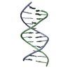
- PDB-1vtd: UNUSUAL HELICAL PACKING IN CRYSTALS OF DNA BEARING A MUTATION HOT SPOT -
+ Open data
- Basic information
| Entry | Database: PDB / ID: 1vtd | ||||||
|---|---|---|---|---|---|---|---|
| Title | UNUSUAL HELICAL PACKING IN CRYSTALS OF DNA BEARING A MUTATION HOT SPOT | ||||||
 Components Components |
| ||||||
 Keywords Keywords | DNA / B-DNA / DOUBLE HELIX | ||||||
| Function / homology | DNA / DNA (> 10) Function and homology information Function and homology information | ||||||
| Biological species | synthetic construct (others) | ||||||
| Method |  X-RAY DIFFRACTION / Resolution: 2.8 Å X-RAY DIFFRACTION / Resolution: 2.8 Å | ||||||
 Authors Authors | Timsit, Y. / Westhof, E. / Fuchs, R.P.P. / Moras, D. | ||||||
 Citation Citation |  Journal: Nature / Year: 1989 Journal: Nature / Year: 1989Title: Unusual helical packing in crystals of DNA bearing a mutation hot spot. Authors: Timsit, Y. / Westhof, E. / Fuchs, R.P. / Moras, D. #1: Journal: J. Mol. Biol. / Year: 1995 Title: Self-fitting and self-modifying properties of the B-DNA molecule. Authors: Timsit, Y. / Moras, D. #2:  Journal: Nature / Year: 1991 Journal: Nature / Year: 1991Title: Base-pairing shift in the major groove of (CA)n tracts by B-DNA crystal structures. Authors: Timsit, Y. / Vilbois, E. / Moras, D. | ||||||
| History |
|
- Structure visualization
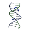
| Structure viewer | Molecule:  Molmil Molmil Jmol/JSmol Jmol/JSmol |
|---|
- Downloads & links
- Download
| PDBx/mmCIF format |  1vtd.cif.gz 1vtd.cif.gz | 21.9 KB | Display |  PDBx/mmCIF format PDBx/mmCIF format |
|---|---|---|---|---|
| PDB format |  pdb1vtd.ent.gz pdb1vtd.ent.gz | 14.1 KB | Display |  PDB format PDB format |
| PDBx/mmJSON format |  1vtd.json.gz 1vtd.json.gz | Tree view |  PDBx/mmJSON format PDBx/mmJSON format | |
| Others |  Other downloads Other downloads |
-Validation report
| Summary document |  1vtd_validation.pdf.gz 1vtd_validation.pdf.gz | 318.7 KB | Display |  wwPDB validaton report wwPDB validaton report |
|---|---|---|---|---|
| Full document |  1vtd_full_validation.pdf.gz 1vtd_full_validation.pdf.gz | 333.6 KB | Display | |
| Data in XML |  1vtd_validation.xml.gz 1vtd_validation.xml.gz | 3.5 KB | Display | |
| Data in CIF |  1vtd_validation.cif.gz 1vtd_validation.cif.gz | 4.4 KB | Display | |
| Arichive directory |  https://data.pdbj.org/pub/pdb/validation_reports/vt/1vtd https://data.pdbj.org/pub/pdb/validation_reports/vt/1vtd ftp://data.pdbj.org/pub/pdb/validation_reports/vt/1vtd ftp://data.pdbj.org/pub/pdb/validation_reports/vt/1vtd | HTTPS FTP |
-Related structure data
| Similar structure data |
|---|
- Links
Links
- Assembly
| Deposited unit | 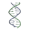
| ||||||||
|---|---|---|---|---|---|---|---|---|---|
| 1 |
| ||||||||
| Unit cell |
|
- Components
| #1: DNA chain | Mass: 3617.371 Da / Num. of mol.: 1 / Source method: obtained synthetically / Source: (synth.) synthetic construct (others) |
|---|---|
| #2: DNA chain | Mass: 3710.401 Da / Num. of mol.: 1 / Source method: obtained synthetically / Source: (synth.) synthetic construct (others) |
-Experimental details
-Experiment
| Experiment | Method:  X-RAY DIFFRACTION X-RAY DIFFRACTION |
|---|
- Sample preparation
| Crystal | Density Matthews: 2.69 Å3/Da / Density % sol: 54.21 % | |||||||||||||||||||||||||||||||||||||||||||||||||
|---|---|---|---|---|---|---|---|---|---|---|---|---|---|---|---|---|---|---|---|---|---|---|---|---|---|---|---|---|---|---|---|---|---|---|---|---|---|---|---|---|---|---|---|---|---|---|---|---|---|---|
| Crystal grow | Temperature: 277 K / Method: vapor diffusion / pH: 6.1 / Details: pH 6.10, VAPOR DIFFUSION, temperature 277.00K | |||||||||||||||||||||||||||||||||||||||||||||||||
| Components of the solutions |
|
-Data collection
| Diffraction | Mean temperature: 273 K |
|---|---|
| Detector | Type: NICOLET / Detector: DIFFRACTOMETER |
| Radiation | Protocol: SINGLE WAVELENGTH / Monochromatic (M) / Laue (L): M / Scattering type: x-ray |
| Radiation wavelength | Relative weight: 1 |
| Reflection | Resolution: 2.8→10 Å / Num. obs: 1584 / Observed criterion σ(F): 1 |
- Processing
| Software | Name: NUCLSQ / Classification: refinement | ||||||||||||
|---|---|---|---|---|---|---|---|---|---|---|---|---|---|
| Refinement | Resolution: 2.8→10 Å / σ(I): 3 /
| ||||||||||||
| Refinement step | Cycle: LAST / Resolution: 2.8→10 Å
|
 Movie
Movie Controller








 PDBj
PDBj


