 Movie
Movie Controller

[English] 日本語
 Yorodumi
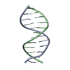
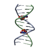
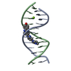
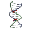
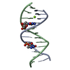
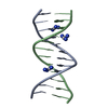
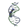
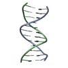
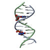
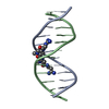
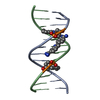
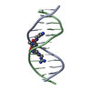
- PDB-1i3t: MOLECULAR AND CRYSTAL STRUCTURE OF D(CGCGAATT(MO4)CGCG): THE WATS... -
+ Open data

- Basic information
| Entry | Database: PDB / ID: 1i3t | ||||||||||||||||||
|---|---|---|---|---|---|---|---|---|---|---|---|---|---|---|---|---|---|---|---|
| Title | MOLECULAR AND CRYSTAL STRUCTURE OF D(CGCGAATT(MO4)CGCG): THE WATSON-CRICK TYPE AND WOBBLE N4-METHOXYCYTIDINE/GUANOSINE BASE PAIRS IN B-DNA | ||||||||||||||||||
 Components Components | 5'-D(* Keywords KeywordsDNA / N4-METHOXYCYTOSINE / DAMAGED DNA / B-DNA / DOUBLE HELIX / DEOXYRIBONUCLEIC ACID | Function / homology | DNA / DNA (> 10) |  Function and homology information Function and homology informationMethod |  X-RAY DIFFRACTION / SYNCHROTRON / MOLECULAR REPLACEMENT / Resolution: 1.6 Å X-RAY DIFFRACTION / SYNCHROTRON / MOLECULAR REPLACEMENT / Resolution: 1.6 Å  Authors AuthorsHossain, M.T. / Hikima, T. / Chatake, T. / Masaru, T. / Sunami, T. / Ueno, Y. / Matsuda, A. / Takenaka, A. |  CitationJournal: J.Biochem.(Tokyo) / Year: 2001 CitationJournal: J.Biochem.(Tokyo) / Year: 2001Title: Crystallographic studies on damaged DNAs: III. N(4)-methoxycytosine can form both Watson-Crick type and wobbled base pairs in a B-form duplex. Authors: Hossain, M.T. / Chatake, T. / Hikima, T. / Tsunoda, M. / Sunami, T. / Ueno, Y. / Matsuda, A. / Takenaka, A. History |
|
- Structure visualization
| Structure viewer | Molecule: MolmilJmol/JSmol |
|---|
- Downloads & links
-Download
| PDBx/mmCIF format | 1i3t.cif.gz | 26.2 KB | Display | PDBx/mmCIF format |
|---|---|---|---|---|
| PDB format | pdb1i3t.ent.gz | 16.8 KB | Display | PDB format |
| PDBx/mmJSON format | 1i3t.json.gz | Tree view | PDBx/mmJSON format | |
| Others |  Other downloads Other downloads |
-Validation report
| Arichive directory | https://data.pdbj.org/pub/pdb/validation_reports/i3/1i3tftp://data.pdbj.org/pub/pdb/validation_reports/i3/1i3t | HTTPS FTP |
|---|
-Related structure data
| Related structure data |  1i47C 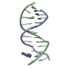 355dS S: Starting model for refinement C: citing same article ( |
|---|---|
| Similar structure data | |
| Other databases |


-Links
 PDBj
PDBj


- Assembly
| Deposited unit | 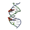
| ||||||||||
|---|---|---|---|---|---|---|---|---|---|---|---|
| 1 |
| ||||||||||
| Unit cell |
|
-Components
| #1: DNA chain | Mass: 3693.418 Da / Num. of mol.: 2 / Source method: obtained synthetically #2: Chemical | ChemComp-MG / |   Mass: 24.305 Da / Num. of mol.: 1 / Source method: obtained synthetically / Formula: Mg Mass: 24.305 Da / Num. of mol.: 1 / Source method: obtained synthetically / Formula: Mg#3: Water | ChemComp-HOH / |  Mass: 18.015 Da / Num. of mol.: 110 / Source method: isolated from a natural source / Formula: H2O Mass: 18.015 Da / Num. of mol.: 110 / Source method: isolated from a natural source / Formula: H2O |
|---|
-Experimental details
-Experiment
| Experiment | Method: X-RAY DIFFRACTION / Number of used crystals: 2 |
|---|
- Sample preparation
| Crystal | Density Matthews: 2.14 Å3/Da / Density % sol: 42.65 % | ||||||||||||||||||||||||||||||
|---|---|---|---|---|---|---|---|---|---|---|---|---|---|---|---|---|---|---|---|---|---|---|---|---|---|---|---|---|---|---|---|
| Crystal grow | Temperature: 277 K / Method: vapor diffusion, hanging drop / pH: 7 Details: MPD, magnesium acetate, spermine, sodium cacodylate. pH 7.0, VAPOR DIFFUSION, HANGING DROP at 277 K | ||||||||||||||||||||||||||||||
| Components of the solutions |
| ||||||||||||||||||||||||||||||
| Crystal grow | *PLUS Temperature: 4 ℃ | ||||||||||||||||||||||||||||||
| Components of the solutions | *PLUS
|
-Data collection
| Diffraction |
| |||||||||||||||
|---|---|---|---|---|---|---|---|---|---|---|---|---|---|---|---|---|
| Diffraction source |
| |||||||||||||||
| Detector |
| |||||||||||||||
| Radiation | Protocol: SINGLE WAVELENGTH / Monochromatic (M) / Laue (L): M / Scattering type: x-ray | |||||||||||||||
| Radiation wavelength | Wavelength: 1 Å / Relative weight: 1 | |||||||||||||||
| Reflection | Resolution: 1.6→100 Å / Num. all: 9068 / Num. obs: 9068 / % possible obs: 97.6 % / Observed criterion σ(I): -3 / Biso Wilson estimate: 13.5 Å2 / Rmerge(I) obs: 0.08 | |||||||||||||||
| Reflection shell | Resolution: 1.6→1.66 Å / Num. unique all: 759 / % possible all: 93.9 | |||||||||||||||
| Reflection | *PLUS Lowest resolution: 100 Å / Rmerge(I) obs: 0.08 | |||||||||||||||
| Reflection shell | *PLUS % possible obs: 93.9 % |

- Processing
| Software |
| |||||||||||||||||||||||||
|---|---|---|---|---|---|---|---|---|---|---|---|---|---|---|---|---|---|---|---|---|---|---|---|---|---|---|
| Refinement | Method to determine structure: MOLECULAR REPLACEMENT Starting model: PDB ENTRY 355D Resolution: 1.6→10 Å / Rfactor Rfree error: 0.008 / Cross valid method: THROUGHOUT / σ(F): 0
| |||||||||||||||||||||||||
| Displacement parameters | Biso mean: 24.5 Å2
| |||||||||||||||||||||||||
| Refine analyze |
| |||||||||||||||||||||||||
| Refinement step | Cycle: LAST / Resolution: 1.6→10 Å
| |||||||||||||||||||||||||
| Refine LS restraints |
| |||||||||||||||||||||||||
| LS refinement shell | Resolution: 1.6→1.66 Å / Rfactor Rfree error: 0.05 / Total num. of bins used: 10
| |||||||||||||||||||||||||
| Software | *PLUS Name: CNS / Version: 1 / Classification: refinement | |||||||||||||||||||||||||
| Refinement | *PLUS Highest resolution: 1.6 Å / Lowest resolution: 10 Å / σ(F): 0 / % reflection Rfree: 10.5 % / Rfactor obs: 0.224 | |||||||||||||||||||||||||
| Solvent computation | *PLUS | |||||||||||||||||||||||||
| Displacement parameters | *PLUS Biso mean: 24.5 Å2 | |||||||||||||||||||||||||
| Refine LS restraints | *PLUS
| |||||||||||||||||||||||||
| LS refinement shell | *PLUS Rfactor Rfree: 0.429 / % reflection Rfree: 8.9 % / Rfactor Rwork: 0.376 |
