 Movie
Movie Controller

[English] 日本語
 Yorodumi
- PDB-1vrr: Crystal structure of the restriction endonuclease BstYI complex w... -
+ Open data

- Basic information
| Entry | Database: PDB / ID: 1vrr | |||||||||
|---|---|---|---|---|---|---|---|---|---|---|
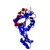
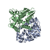
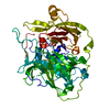
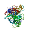
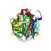
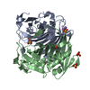
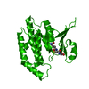
| Title | Crystal structure of the restriction endonuclease BstYI complex with DNA | |||||||||
 Components Components |
| |||||||||
 Keywords Keywords | hydrolase/DNA / Enzyme-DNA complex / double helix / hydrolase-DNA COMPLEX | |||||||||
| Function / homology |  Function and homology information Function and homology informationtype II site-specific deoxyribonuclease activity / DNA restriction-modification system / magnesium ion binding / DNA binding / identical protein binding Similarity search - Function | |||||||||
| Biological species |   Geobacillus stearothermophilus (bacteria) Geobacillus stearothermophilus (bacteria) | |||||||||
| Method |  X-RAY DIFFRACTION / SYNCHROTRON / MAD / Resolution: 2.7 Å X-RAY DIFFRACTION / SYNCHROTRON / MAD / Resolution: 2.7 Å | |||||||||
 Authors Authors | Townson, S.A. / Samuelson, J.C. / Xu, S.Y. / Aggarwal, A.K. | |||||||||
 Citation Citation | Journal: Structure / Year: 2005 Title: Implications for Switching Restriction Enzyme Specificities from the Structure of BstYI Bound to a BglII DNA Sequence. Authors: Townson, S.A. / Samuelson, J.C. / Xu, S.Y. / Aggarwal, A.K. | |||||||||
| History |
|
- Structure visualization
| Structure viewer | Molecule: MolmilJmol/JSmol |
|---|
- Downloads & links
-Download
| PDBx/mmCIF format | 1vrr.cif.gz | 108.8 KB | Display | PDBx/mmCIF format |
|---|---|---|---|---|
| PDB format | pdb1vrr.ent.gz | 81.4 KB | Display | PDB format |
| PDBx/mmJSON format | 1vrr.json.gz | Tree view | PDBx/mmJSON format | |
| Others |  Other downloads Other downloads |
-Validation report
| Arichive directory | https://data.pdbj.org/pub/pdb/validation_reports/vr/1vrrftp://data.pdbj.org/pub/pdb/validation_reports/vr/1vrr | HTTPS FTP |
|---|
-Related structure data
| Related structure data | |
|---|---|
| Similar structure data |


-Links
 PDBj
PDBj


- Assembly
| Deposited unit | 
| ||||||||
|---|---|---|---|---|---|---|---|---|---|
| 1 |
| ||||||||
| Unit cell |
| ||||||||
| Details | The biological assembly is composed of the BstYI dimer (chains A and B) and a 14-mer DNA duplex (chains C and D) |
-Components
| #1: DNA chain | Mass: 4277.828 Da / Num. of mol.: 2 / Source method: obtained synthetically #2: Protein | Mass: 23222.674 Da / Num. of mol.: 2 Source method: isolated from a genetically manipulated source Details: Type II Restriction Endonuclease BstYI Source: (gene. exp.) Geobacillus stearothermophilus (bacteria)Gene: bstYIR / Plasmid: pACYC,pCEF8,pET21at / Production host: References: GenBank: 28864483, UniProt: Q84AF2*PLUS, type II site-specific deoxyribonuclease #3: Water | ChemComp-HOH / |  Mass: 18.015 Da / Num. of mol.: 104 / Source method: isolated from a natural source / Formula: H2O Mass: 18.015 Da / Num. of mol.: 104 / Source method: isolated from a natural source / Formula: H2O |
|---|
-Experimental details
-Experiment
| Experiment | Method: X-RAY DIFFRACTION / Number of used crystals: 1 |
|---|
- Sample preparation
| Crystal | Density Matthews: 3.032 Å3/Da / Density % sol: 57.87 % | ||||||||||||||||||||||||||||
|---|---|---|---|---|---|---|---|---|---|---|---|---|---|---|---|---|---|---|---|---|---|---|---|---|---|---|---|---|---|
| Crystal grow | Temperature: 293 K / Method: vapor diffusion, hanging drop / pH: 6.5 Details: 1.0 M sodium citrate, 0.1 M sodium cacodylate, 1.5% 1,2,3-heptanetriol, pH 6.5, VAPOR DIFFUSION, HANGING DROP, temperature 293K | ||||||||||||||||||||||||||||
| Components of the solutions |
|
-Data collection
| Diffraction | Mean temperature: 110 K | |||||||||
|---|---|---|---|---|---|---|---|---|---|---|
| Diffraction source | Source: SYNCHROTRON / Site: NSLS  / Beamline: X12C / Wavelength: 0.979,0.968 / Beamline: X12C / Wavelength: 0.979,0.968 | |||||||||
| Detector | Type: ENRAF-NONIUS FAST / Detector: DIFFRACTOMETER / Date: Sep 3, 2003 | |||||||||
| Radiation | Monochromator: channel-cut crystal monochromator with toroidal mirrors Protocol: MAD / Monochromatic (M) / Laue (L): M / Scattering type: x-ray | |||||||||
| Radiation wavelength |
| |||||||||
| Reflection | Resolution: 2.7→50 Å / Num. obs: 35479 / % possible obs: 67.9 % / Observed criterion σ(F): 1 / Observed criterion σ(I): 1 / Redundancy: 6.2 % | |||||||||
| Reflection shell | Highest resolution: 2.7 Å / % possible all: 10 |
- Processing
| Software |
| ||||||||||||||||||||
|---|---|---|---|---|---|---|---|---|---|---|---|---|---|---|---|---|---|---|---|---|---|
| Refinement | Method to determine structure: MAD / Resolution: 2.7→50 Å / Cross valid method: THROUGHOUT / σ(F): 1 / Stereochemistry target values: Engh & Huber
| ||||||||||||||||||||
| Refine analyze |
| ||||||||||||||||||||
| Refinement step | Cycle: LAST / Resolution: 2.7→50 Å
|
