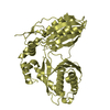
 Movie
Movie Controller


+ Open data

- Basic information
| Entry | Database: PDB / ID: 1sdo | ||||||
|---|---|---|---|---|---|---|---|
| Title | Crystal Structure of Restriction Endonuclease BstYI | ||||||
 Components Components | BstYI | ||||||
 Keywords Keywords | HYDROLASE / Restriction Endonuclease | ||||||
| Function / homology |  Function and homology information Function and homology informationtype II site-specific deoxyribonuclease activity / DNA restriction-modification system / magnesium ion binding / DNA binding / identical protein binding Similarity search - Function | ||||||
| Biological species |   Geobacillus stearothermophilus (bacteria) Geobacillus stearothermophilus (bacteria) | ||||||
| Method |  X-RAY DIFFRACTION / SYNCHROTRON / MAD / Resolution: 1.85 Å X-RAY DIFFRACTION / SYNCHROTRON / MAD / Resolution: 1.85 Å | ||||||
 Authors Authors | Townson, S.A. / Samuelson, J.C. / Vanamee, E.S. / Edwards, T.A. / Escalante, C.R. / Xu, S.Y. / Aggarwal, A.K. | ||||||
 Citation Citation | Journal: J.Mol.Biol. / Year: 2004 Title: Crystal Structure of BstYI at 1.85 A Resolution: A Thermophilic Restriction Endonuclease with Overlapping Specificities to BamHI and BglII Authors: Townson, S.A. / Samuelson, J.C. / Vanamee, E.S. / Edwards, T.A. / Escalante, C.R. / Xu, S.Y. / Aggarwal, A.K. | ||||||
| History |
|
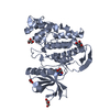
- Structure visualization
| Structure viewer | Molecule: MolmilJmol/JSmol |
|---|
- Downloads & links
-Download
| PDBx/mmCIF format | 1sdo.cif.gz | 50.3 KB | Display | PDBx/mmCIF format |
|---|---|---|---|---|
| PDB format | pdb1sdo.ent.gz | 36.6 KB | Display | PDB format |
| PDBx/mmJSON format | 1sdo.json.gz | Tree view | PDBx/mmJSON format | |
| Others |  Other downloads Other downloads |
-Validation report
| Arichive directory | https://data.pdbj.org/pub/pdb/validation_reports/sd/1sdoftp://data.pdbj.org/pub/pdb/validation_reports/sd/1sdo | HTTPS FTP |
|---|
-Related structure data
| Similar structure data |
|---|


-Links
 PDBj
PDBj


- Assembly
| Deposited unit | 
| ||||||||
|---|---|---|---|---|---|---|---|---|---|
| 1 | 
| ||||||||
| Unit cell |
| ||||||||
| Details | This entry contains the crystallographic asymmetric unit which consists of a monomer (1 chain). The biological dimer is generated by the operations: Rotation Matrix -1.00000 0.00002 0.00000 0.00002 1.00000 -0.00004 0.00000 -0.00004 -1.00000 Translation 69.89 0.0094 78.9707 |
-Components
| #1: Protein | Mass: 23222.674 Da / Num. of mol.: 1 Source method: isolated from a genetically manipulated source Source: (gene. exp.) Geobacillus stearothermophilus (bacteria)Gene: BSTYIR / Production host: References: UniProt: Q84AF2, type II site-specific deoxyribonuclease |
|---|---|
| #2: Water | ChemComp-HOH /  Mass: 18.015 Da / Num. of mol.: 90 / Source method: isolated from a natural source / Formula: H2O Mass: 18.015 Da / Num. of mol.: 90 / Source method: isolated from a natural source / Formula: H2O |
-Experimental details
-Experiment
| Experiment | Method: X-RAY DIFFRACTION / Number of used crystals: 2 |
|---|
- Sample preparation
| Crystal | Density Matthews: 2.28 Å3/Da / Density % sol: 45.94 % |
|---|---|
| Crystal grow | Temperature: 298 K / Method: vapor diffusion, hanging drop / pH: 7.5 Details: 30% PEG-400, 0.1M HEPES, 200mM Calcium chloride, pH 7.5, VAPOR DIFFUSION, HANGING DROP, temperature 298K |
-Data collection
| Diffraction | Mean temperature: 100 K | |||||||||
|---|---|---|---|---|---|---|---|---|---|---|
| Diffraction source | Source: SYNCHROTRON / Site: APS  / Beamline: 19-ID / Wavelength: 0.973, 0.9282 / Beamline: 19-ID / Wavelength: 0.973, 0.9282 | |||||||||
| Detector | Type: SBC-2 / Detector: CCD / Date: Jan 1, 2002 | |||||||||
| Radiation | Protocol: MAD / Monochromatic (M) / Laue (L): M / Scattering type: x-ray | |||||||||
| Radiation wavelength |
| |||||||||
| Reflection | Resolution: 1.85→50 Å / Num. all: 34966 / Num. obs: 33604 / % possible obs: 98.9 % / Observed criterion σ(F): 2 / Observed criterion σ(I): 0 / Rmerge(I) obs: 0.092 / Rsym value: 0.085 / Net I/σ(I): 17.4 |
- Processing
| Software |
| |||||||||||||||||||||||||
|---|---|---|---|---|---|---|---|---|---|---|---|---|---|---|---|---|---|---|---|---|---|---|---|---|---|---|
| Refinement | Method to determine structure: MAD / Resolution: 1.85→50 Å / Cross valid method: THROUGHOUT / σ(F): 2 / σ(I): 0 / Stereochemistry target values: Engh & Huber
| |||||||||||||||||||||||||
| Displacement parameters | Biso mean: 39.6 Å2
| |||||||||||||||||||||||||
| Refine analyze |
| |||||||||||||||||||||||||
| Refinement step | Cycle: LAST / Resolution: 1.85→50 Å
| |||||||||||||||||||||||||
| Refine LS restraints |
|
