 Movie
Movie Controller

[English] 日本語
 Yorodumi
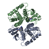
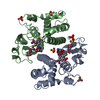
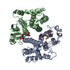
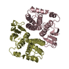
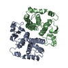
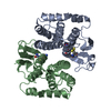
- PDB-1gsu: AN AVIAN CLASS-MU GLUTATHIONE S-TRANSFERASE, CGSTM1-1 AT 1.94 ANG... -
+ Open data

- Basic information
| Entry | Database: PDB / ID: 1gsu | ||||||
|---|---|---|---|---|---|---|---|
| Title | AN AVIAN CLASS-MU GLUTATHIONE S-TRANSFERASE, CGSTM1-1 AT 1.94 ANGSTROM RESOLUTION | ||||||
 Components Components | CLASS-MU GLUTATHIONE S-TRANSFERASE | ||||||
 Keywords Keywords | DETOXIFICATION ENZYME / GLUTATHIONE S-TRANSFERASE / S-HEXYL GLUTATHIONE | ||||||
| Function / homology |  Function and homology information Function and homology informationGlutathione conjugation / Biosynthesis of maresin conjugates in tissue regeneration (MCTR) / Azathioprine ADME / Paracetamol ADME / glutathione transferase / glutathione transferase activity / lipid metabolic process / glutathione metabolic process / identical protein binding / cytoplasm Similarity search - Function | ||||||
| Biological species |  | ||||||
| Method |  X-RAY DIFFRACTION / Resolution: 1.94 Å X-RAY DIFFRACTION / Resolution: 1.94 Å | ||||||
 Authors Authors | Sun, Y.-J. / Kuan, C. / Tam, M.F. / Hsiao, C.-D. | ||||||
 Citation Citation | Journal: J.Mol.Biol. / Year: 1998 Title: The three-dimensional structure of an avian class-mu glutathione S-transferase, cGSTM1-1 at 1.94 A resolution. Authors: Sun, Y.J. / Kuan, I.C. / Tam, M.F. / Hsiao, C.D. #1: Journal: Biochemistry / Year: 1994Title: Structure and Function of the Xenobiotic Substrate Binding Site of a Glutathione S-Transferase as Revealed by X-Ray Crystallographic Analysis of Product Complexes with the Diastereomers of 9- ...Title: Structure and Function of the Xenobiotic Substrate Binding Site of a Glutathione S-Transferase as Revealed by X-Ray Crystallographic Analysis of Product Complexes with the Diastereomers of 9-(S-Glutathionyl)-10-Hydroxy-9,10-Dihydrophenanthrene Authors: Ji, X. / Johnson, W.W. / Sesay, M.A. / Dickert, L. / Prasad, S.M. / Ammon, H.L. / Armstrong, R.N. / Gilliland, G.L. #2: Journal: J.Mol.Biol. / Year: 1994Title: Crystal Structure of Human Class Mu Glutathione Transferase Gstm2-2. Effects of Lattice Packing on Conformational Heterogeneity Authors: Raghunathan, S. / Chandross, R.J. / Kretsinger, R.H. / Allison, T.J. / Penington, C.J. / Rule, G.S. #3: Journal: Biochemistry / Year: 1992Title: The Three-Dimensional Structure of a Glutathione S-Transferase from the Mu Gene Class. Structural Analysis of the Binary Complex of Isoenzyme 3-3 and Glutathione at 2.2-A Resolution Authors: Ji, X. / Zhang, P. / Armstrong, R.N. / Gilliland, G.L. | ||||||
| History |
|
- Structure visualization
| Structure viewer | Molecule: MolmilJmol/JSmol |
|---|
- Downloads & links
-Download
| PDBx/mmCIF format | 1gsu.cif.gz | 100.7 KB | Display | PDBx/mmCIF format |
|---|---|---|---|---|
| PDB format | pdb1gsu.ent.gz | 79.1 KB | Display | PDB format |
| PDBx/mmJSON format | 1gsu.json.gz | Tree view | PDBx/mmJSON format | |
| Others |  Other downloads Other downloads |
-Validation report
| Arichive directory | https://data.pdbj.org/pub/pdb/validation_reports/gs/1gsuftp://data.pdbj.org/pub/pdb/validation_reports/gs/1gsu | HTTPS FTP |
|---|
-Related structure data
| Similar structure data |
|---|


-Links
 PDBj
PDBj

- Assembly
| Deposited unit | 
| ||||||||
|---|---|---|---|---|---|---|---|---|---|
| 1 |
| ||||||||
| Unit cell |
|
-Components
| #1: Protein | Mass: 25794.621 Da / Num. of mol.: 2 Source method: isolated from a genetically manipulated source Source: (gene. exp.)  #2: Chemical | 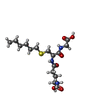  Mass: 392.491 Da / Num. of mol.: 2 / Source method: obtained synthetically / Formula: C16H30N3O6S Mass: 392.491 Da / Num. of mol.: 2 / Source method: obtained synthetically / Formula: C16H30N3O6S#3: Water | ChemComp-HOH / |  Mass: 18.015 Da / Num. of mol.: 57 / Source method: isolated from a natural source / Formula: H2O Mass: 18.015 Da / Num. of mol.: 57 / Source method: isolated from a natural source / Formula: H2O |
|---|
-Experimental details
-Experiment
| Experiment | Method: X-RAY DIFFRACTION |
|---|
- Sample preparation
| Crystal | Density Matthews: 2.45 Å3/Da / Density % sol: 50 % | ||||||||||||||||||
|---|---|---|---|---|---|---|---|---|---|---|---|---|---|---|---|---|---|---|---|
| Crystal grow | *PLUS Method: vapor diffusion, hanging drop | ||||||||||||||||||
| Components of the solutions | *PLUS
|
-Data collection
| Diffraction source | Wavelength: 1.5418 |
|---|---|
| Detector | Type: RIGAKU RAXIS II / Detector: IMAGE PLATE / Date: Jan 25, 1996 |
| Radiation | Monochromatic (M) / Laue (L): M / Scattering type: x-ray |
| Radiation wavelength | Wavelength: 1.5418 Å / Relative weight: 1 |
| Reflection | Num. obs: 32795 / % possible obs: 86.7 % / Observed criterion σ(I): 0 / Redundancy: 4 % / Rmerge(I) obs: 0.091 |
| Reflection | *PLUS Highest resolution: 1.94 Å / Num. measured all: 129695 |
- Processing
| Software |
| ||||||||||||||||||||||||||||||||||||||||||||||||||||||||||||
|---|---|---|---|---|---|---|---|---|---|---|---|---|---|---|---|---|---|---|---|---|---|---|---|---|---|---|---|---|---|---|---|---|---|---|---|---|---|---|---|---|---|---|---|---|---|---|---|---|---|---|---|---|---|---|---|---|---|---|---|---|---|
| Refinement | Resolution: 1.94→6 Å / σ(F): 2
| ||||||||||||||||||||||||||||||||||||||||||||||||||||||||||||
| Displacement parameters | Biso mean: 22.6 Å2 | ||||||||||||||||||||||||||||||||||||||||||||||||||||||||||||
| Refine analyze | Luzzati coordinate error obs: 0.3 Å | ||||||||||||||||||||||||||||||||||||||||||||||||||||||||||||
| Refinement step | Cycle: LAST / Resolution: 1.94→6 Å
| ||||||||||||||||||||||||||||||||||||||||||||||||||||||||||||
| Refine LS restraints |
|
