- PDB-1vr8: Crystal structure of GTP binding regulator (TM1622) from Thermoto... -
+
Open data
ID or keywords:
Loading...
-
Basic information
Entry
Database: PDB / ID: 1vr8
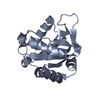
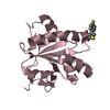
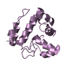
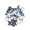
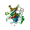
Title
Crystal structure of GTP binding regulator (TM1622) from Thermotoga Maritima at 1.75 A resolution
Components
GTP binding regulator
Keywords
SIGNALING PROTEIN / TM1622 / GTP BINDING REGULATOR / Structural Genomics / Joint Center for Structural Genomics / JCSG / Protein Structure Initiative / PSI
Function / homology
Function and homology information
TM1622-like / Protein of unknown function DUF3242 / TM1622-like superfamily / Protein of unknown function (DUF3242) / Protein Transport Mog1p; Chain A / Prokaryotic membrane lipoprotein lipid attachment site profile. / 3-Layer(aba) Sandwich / Alpha Beta Similarity search - Domain/homology
SEQUENCE THE CONSTRUCT EXPRESSED COMPRISED AN N-TERMINAL PURIFICATION TAG [MGSDKIHHHHHH] FOLLOWED ...SEQUENCE THE CONSTRUCT EXPRESSED COMPRISED AN N-TERMINAL PURIFICATION TAG [MGSDKIHHHHHH] FOLLOWED BY RESIDUES 25-154 OF THE PREDICTED TM1622 GENE PRODUCT. IN ORDER TO REMOVE A PREDICTED TRANSMEMBRANE HELIX, THE FIRST 24 RESIDUES WERE NOT INCLUDED IN THE CONSTRUCT.
Resolution: 1.75→29.31 Å / Num. obs: 16407 / % possible obs: 93.2 % / Redundancy: 4.4 % / Biso Wilson estimate: 25.41 Å2 / Rsym value: 0.068 / Net I/σ(I): 14.4
Reflection shell
Resolution: 1.75→1.85 Å / Redundancy: 1.7 % / Mean I/σ(I) obs: 2.3 / Num. unique all: 1586 / Rsym value: 0.339 / % possible all: 63.9
-
Processing
Software
Name
Version
Classification
MOSFLM
datareduction
SCALA
5
datascaling
SHELXD
phasing
autoSHARP
phasing
REFMAC
5.2.0005
refinement
CCP4
(SCALA)
datascaling
Refinement
Method to determine structure: MAD / Resolution: 1.75→29.31 Å / Cor.coef. Fo:Fc: 0.966 / Cor.coef. Fo:Fc free: 0.949 / SU B: 3.433 / SU ML: 0.056 / TLS residual ADP flag: LIKELY RESIDUAL / Cross valid method: THROUGHOUT / ESU R: 0.096 / ESU R Free: 0.096 Stereochemistry target values: MAXIMUM LIKELIHOOD WITH PHASES Details: 1. HYDROGENS HAVE BEEN ADDED IN THE RIDING POSITIONS. 2. LOOP 93-96 IS DISORDERED. THERE IS SOME EXTRA UNKNOWN DENSITY NEARBY WHICH IS NOT MODELED AS WELL.
Rfactor
Num. reflection
% reflection
Selection details
Rfree
0.18179
832
5.1 %
RANDOM
Rwork
0.1486
-
-
-
obs
0.15019
15549
93.16 %
-
Solvent computation
Ion probe radii: 0.8 Å / Shrinkage radii: 0.8 Å / VDW probe radii: 1.2 Å / Solvent model: BABINET MODEL WITH MASK
Displacement parameters
Biso mean: 23.241 Å2
Baniso -1
Baniso -2
Baniso -3
1-
-0.02 Å2
-0.01 Å2
0 Å2
2-
-
-0.02 Å2
0 Å2
3-
-
-
0.04 Å2
Refinement step
Cycle: LAST / Resolution: 1.75→29.31 Å
Protein
Nucleic acid
Ligand
Solvent
Total
Num. atoms
1098
0
24
172
1294
Refine LS restraints
Refine-ID
Type
Dev ideal
Dev ideal target
Number
X-RAY DIFFRACTION
r_bond_refined_d
0.014
0.022
1160
X-RAY DIFFRACTION
r_bond_other_d
0.001
0.02
1054
X-RAY DIFFRACTION
r_angle_refined_deg
1.49
1.931
1560
X-RAY DIFFRACTION
r_angle_other_deg
0.738
3
2433
X-RAY DIFFRACTION
r_dihedral_angle_1_deg
5.627
5
135
X-RAY DIFFRACTION
r_dihedral_angle_2_deg
33.444
22.885
52
X-RAY DIFFRACTION
r_dihedral_angle_3_deg
12.467
15
197
X-RAY DIFFRACTION
r_dihedral_angle_4_deg
10.793
15
6
X-RAY DIFFRACTION
r_chiral_restr
0.09
0.2
164
X-RAY DIFFRACTION
r_gen_planes_refined
0.006
0.02
1244
X-RAY DIFFRACTION
r_gen_planes_other
0.001
0.02
250
X-RAY DIFFRACTION
r_nbd_refined
0.195
0.2
201
X-RAY DIFFRACTION
r_nbd_other
0.171
0.2
1049
X-RAY DIFFRACTION
r_nbtor_other
0.085
0.2
705
X-RAY DIFFRACTION
r_xyhbond_nbd_refined
0.15
0.2
115
X-RAY DIFFRACTION
r_symmetry_vdw_refined
0.164
0.2
10
X-RAY DIFFRACTION
r_symmetry_vdw_other
0.245
0.2
39
X-RAY DIFFRACTION
r_symmetry_hbond_refined
0.225
0.2
26
X-RAY DIFFRACTION
r_mcbond_it
2.299
3
716
X-RAY DIFFRACTION
r_mcbond_other
0.554
3
278
X-RAY DIFFRACTION
r_mcangle_it
2.897
5
1092
X-RAY DIFFRACTION
r_scbond_it
3.196
5
562
X-RAY DIFFRACTION
r_scangle_it
4.192
5
467
X-RAY DIFFRACTION
r_nbtor_refined
0.187
0.2
548
LS refinement shell
Resolution: 1.75→1.797 Å / Total num. of bins used: 20
Rfactor
Num. reflection
% reflection
Rfree
0.252
37
5.52 %
Rwork
0.224
633
-
obs
-
-
53.09 %
Refinement TLS params.
Method: refined / Origin x: 36.8774 Å / Origin y: -12.4241 Å / Origin z: -16.2932 Å
11
12
13
21
22
23
31
32
33
T
-0.167 Å2
0.0323 Å2
-0.0125 Å2
-
-0.1722 Å2
0.0026 Å2
-
-
-0.1955 Å2
L
1.0805 °2
0.1108 °2
0.1823 °2
-
3.3665 °2
0.0302 °2
-
-
1.2724 °2
S
-0.0152 Å °
0.0314 Å °
0.0078 Å °
-0.1138 Å °
0.0238 Å °
-0.0872 Å °
0.0083 Å °
0.046 Å °
-0.0086 Å °
Refinement TLS group
Selection: ALL
+
About Yorodumi
-
News
-
Feb 9, 2022. New format data for meta-information of EMDB entries
New format data for meta-information of EMDB entries
Version 3 of the EMDB header file is now the official format.
The previous official version 1.9 will be removed from the archive.
In the structure databanks used in Yorodumi, some data are registered as the other names, "COVID-19 virus" and "2019-nCoV". Here are the details of the virus and the list of structure data.
Jan 31, 2019. EMDB accession codes are about to change! (news from PDBe EMDB page)
EMDB accession codes are about to change! (news from PDBe EMDB page)
The allocation of 4 digits for EMDB accession codes will soon come to an end. Whilst these codes will remain in use, new EMDB accession codes will include an additional digit and will expand incrementally as the available range of codes is exhausted. The current 4-digit format prefixed with “EMD-” (i.e. EMD-XXXX) will advance to a 5-digit format (i.e. EMD-XXXXX), and so on. It is currently estimated that the 4-digit codes will be depleted around Spring 2019, at which point the 5-digit format will come into force.
The EM Navigator/Yorodumi systems omit the EMD- prefix.
Related info.:Q: What is EMD? / ID/Accession-code notation in Yorodumi/EM Navigator
Yorodumi is a browser for structure data from EMDB, PDB, SASBDB, etc.
This page is also the successor to EM Navigator detail page, and also detail information page/front-end page for Omokage search.
The word "yorodu" (or yorozu) is an old Japanese word meaning "ten thousand". "mi" (miru) is to see.
Related info.:EMDB / PDB / SASBDB / Comparison of 3 databanks / Yorodumi Search / Aug 31, 2016. New EM Navigator & Yorodumi / Yorodumi Papers / Jmol/JSmol / Function and homology information / Changes in new EM Navigator and Yorodumi
 Movie
Movie Controller

 Yorodumi
 Open data
 Basic information
 Components
 Keywords
 Function and homology information
 Thermotoga maritima (bacteria)
 X-RAY DIFFRACTION /
  Authors
 Citation
 Structure visualization
 Downloads & links
 Other downloads


 PDBj
PDBj


 Assembly
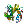

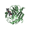
 Mass: 42.020 Da / Num. of mol.: 2 / Source method: obtained synthetically / Formula: N3
 Mass: 92.094 Da / Num. of mol.: 3 / Source method: obtained synthetically / Formula: C3H8O3
 Mass: 18.015 Da / Num. of mol.: 172 / Source method: isolated from a natural source / Formula: H2O
 Sample preparation
 / Beamline: 8.2.1 / Wavelength: 0.99186, 0.97951, 0.97933
 Processing