 Movie
Movie Controller


+ Open data

- Basic information
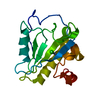
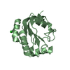
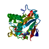
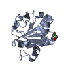
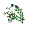
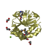
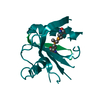
| Entry | Database: PDB / ID: 3hmb | ||||||
|---|---|---|---|---|---|---|---|
| Title | Mutant endolysin from Bacillus subtilis | ||||||
 Components Components | N-acetylmuramoyl-L-alanine amidase xlyA | ||||||
 Keywords Keywords | HYDROLASE / endolysin / amidase / Cell wall biogenesis/degradation / Competence / Secreted / Sporulation | ||||||
| Function / homology |  Function and homology information Function and homology informationN-acetylmuramoyl-L-alanine amidase / establishment of competence for transformation / N-acetylmuramoyl-L-alanine amidase activity / peptidoglycan turnover / sporulation resulting in formation of a cellular spore / peptidoglycan catabolic process / cell wall organization / extracellular region Similarity search - Function | ||||||
| Biological species |  | ||||||
| Method |  X-RAY DIFFRACTION / MOLECULAR REPLACEMENT / Resolution: 2.7 Å X-RAY DIFFRACTION / MOLECULAR REPLACEMENT / Resolution: 2.7 Å | ||||||
 Authors Authors | Low, L.Y. / Liddington, R. | ||||||
 Citation Citation | Journal: J.Biol.Chem. / Year: 2011 Title: Role of net charge on catalytic domain and influence of cell wall binding domain on bactericidal activity, specificity, and host range of phage lysins. Authors: Low, L.Y. / Yang, C. / Perego, M. / Osterman, A. / Liddington, R. | ||||||
| History |
|
- Structure visualization
| Structure viewer | Molecule: MolmilJmol/JSmol |
|---|
- Downloads & links
-Download
| PDBx/mmCIF format | 3hmb.cif.gz | 102.4 KB | Display | PDBx/mmCIF format |
|---|---|---|---|---|
| PDB format | pdb3hmb.ent.gz | 78.3 KB | Display | PDB format |
| PDBx/mmJSON format | 3hmb.json.gz | Tree view | PDBx/mmJSON format | |
| Others |  Other downloads Other downloads |
-Validation report
| Arichive directory | https://data.pdbj.org/pub/pdb/validation_reports/hm/3hmbftp://data.pdbj.org/pub/pdb/validation_reports/hm/3hmb | HTTPS FTP |
|---|
-Related structure data
| Related structure data | 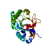 3hmcC  3rdrC  3hma C: citing same article ( S: Starting model for refinement |
|---|---|
| Similar structure data |


-Links
 PDBj
PDBj

- Assembly
| Deposited unit | 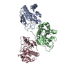
| ||||||||
|---|---|---|---|---|---|---|---|---|---|
| 1 | 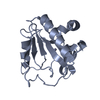
| ||||||||
| 2 | 
| ||||||||
| 3 | 
| ||||||||
| Unit cell |
|
-Components
| #1: Protein | Mass: 17222.312 Da / Num. of mol.: 3 / Mutation: D7K, T22K, L24K, T63K, T145K Source method: isolated from a genetically manipulated source Source: (gene. exp.) References: UniProt: P39800, N-acetylmuramoyl-L-alanine amidase #2: Chemical |   Mass: 65.409 Da / Num. of mol.: 3 / Source method: obtained synthetically / Formula: Zn Mass: 65.409 Da / Num. of mol.: 3 / Source method: obtained synthetically / Formula: Zn#3: Water | ChemComp-HOH / |  Mass: 18.015 Da / Num. of mol.: 149 / Source method: isolated from a natural source / Formula: H2O Mass: 18.015 Da / Num. of mol.: 149 / Source method: isolated from a natural source / Formula: H2O |
|---|
-Experimental details
-Experiment
| Experiment | Method: X-RAY DIFFRACTION / Number of used crystals: 1 |
|---|
- Sample preparation
| Crystal | Density Matthews: 4.37 Å3/Da / Density % sol: 71.88 % |
|---|
-Data collection
| Diffraction | Mean temperature: 100 K |
|---|---|
| Diffraction source | Source: ROTATING ANODE / Type: RIGAKU FR-E SUPERBRIGHT / Wavelength: 1.54 Å |
| Detector | Type: RIGAKU RAXIS IV / Detector: IMAGE PLATE / Date: Feb 7, 2005 |
| Radiation | Protocol: SINGLE WAVELENGTH / Monochromatic (M) / Laue (L): M / Scattering type: x-ray |
| Radiation wavelength | Wavelength: 1.54 Å / Relative weight: 1 |
| Reflection | Resolution: 2.7→40 Å / Num. obs: 25390 / % possible obs: 99.9 % / Observed criterion σ(F): 2 / Redundancy: 6.4 % / Rmerge(I) obs: 0.2 / Net I/σ(I): 10 |
| Reflection shell | Resolution: 2.7→2.85 Å / % possible all: 100 |
- Processing
| Software |
| ||||||||||||||||||||
|---|---|---|---|---|---|---|---|---|---|---|---|---|---|---|---|---|---|---|---|---|---|
| Refinement | Method to determine structure: MOLECULAR REPLACEMENT Starting model: pdb entry 3HMA 3hma Resolution: 2.7→40 Å / Isotropic thermal model: isotropic
| ||||||||||||||||||||
| Refinement step | Cycle: LAST / Resolution: 2.7→40 Å
|
