 Movie
Movie Controller

[English] 日本語
 Yorodumi
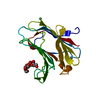
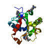
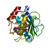
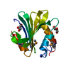
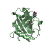
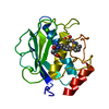
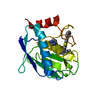
- PDB-1xiz: Structural Genomics, The crystal structure of domain IIA of putat... -
+ Open data

- Basic information
| Entry | Database: PDB / ID: 1xiz | ||||||
|---|---|---|---|---|---|---|---|
| Title | Structural Genomics, The crystal structure of domain IIA of putative phosphotransferase system specific for mannitol/fructose from Salmonella typhimurium | ||||||
 Components Components | putative phosphotransferase system mannitol/fructose-specific IIA domain | ||||||
 Keywords Keywords | STRUCTURAL GENOMICS / UNKNOWN FUNCTION / IIA domain / protein structure initiative / Midwest Center for Structural Genomics / MCSG / PSI | ||||||
| Function / homology |  Function and homology information Function and homology information: / PTS EIIA type-2 domain / Phosphoenolpyruvate-dependent sugar phosphotransferase system, EIIA 2 / PTS_EIIA type-2 domain profile. / Mannitol-specific EII; Chain A / Mannitol-specific EII; Chain A / Phosphotransferase/anion transporter / 3-Layer(aba) Sandwich / Alpha Beta Similarity search - Domain/homology | ||||||
| Biological species |  Salmonella typhimurium (bacteria) Salmonella typhimurium (bacteria) | ||||||
| Method |  X-RAY DIFFRACTION / SYNCHROTRON / MAD / Resolution: 2 Å X-RAY DIFFRACTION / SYNCHROTRON / MAD / Resolution: 2 Å | ||||||
 Authors Authors | Zhang, R. / Joachimiak, G. / Otwinowski, Z. / Collart, F. / Joachimiak, A. / Midwest Center for Structural Genomics (MCSG) | ||||||
 Citation Citation | Journal: To be Published Title: The crystal structure of domain IIA of putative phosphotransferase system specific for mannitol/fructose from Salmonella typhimurium Authors: Zhang, R. / Joachimiak, G. / Otwinowski, Z. / Collart, F. / Joachimiak, A. | ||||||
| History |
|
- Structure visualization
| Structure viewer | Molecule: MolmilJmol/JSmol |
|---|
- Downloads & links
-Download
| PDBx/mmCIF format | 1xiz.cif.gz | 74.8 KB | Display | PDBx/mmCIF format |
|---|---|---|---|---|
| PDB format | pdb1xiz.ent.gz | 56.4 KB | Display | PDB format |
| PDBx/mmJSON format | 1xiz.json.gz | Tree view | PDBx/mmJSON format | |
| Others |  Other downloads Other downloads |
-Validation report
| Arichive directory | https://data.pdbj.org/pub/pdb/validation_reports/xi/1xizftp://data.pdbj.org/pub/pdb/validation_reports/xi/1xiz | HTTPS FTP |
|---|
-Related structure data
| Similar structure data | |
|---|---|
| Other databases |


-Links
 PDBj
PDBj- Assembly
| Deposited unit | 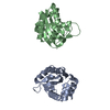
| ||||||||
|---|---|---|---|---|---|---|---|---|---|
| 1 | 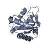
| ||||||||
| 2 | 
| ||||||||
| Unit cell |
|
-Components
| #1: Protein | Mass: 18024.443 Da / Num. of mol.: 2 Source method: isolated from a genetically manipulated source Source: (gene. exp.) Salmonella typhimurium (bacteria) / Plasmid: PDM68 / Species (production host): Escherichia coli / Production host: #2: Water | ChemComp-HOH / |  Mass: 18.015 Da / Num. of mol.: 164 / Source method: isolated from a natural source / Formula: H2O Mass: 18.015 Da / Num. of mol.: 164 / Source method: isolated from a natural source / Formula: H2O |
|---|
-Experimental details
-Experiment
| Experiment | Method: X-RAY DIFFRACTION / Number of used crystals: 1 |
|---|
- Sample preparation
| Crystal | Density Matthews: 2.1 Å3/Da / Density % sol: 39.3 % |
|---|---|
| Crystal grow | Temperature: 298 K / Method: vapor diffusion, sitting drop / pH: 7 Details: 2.4M Sodium Malonate, pH 7.0, VAPOR DIFFUSION, SITTING DROP, temperature 298K |
-Data collection
| Diffraction | Mean temperature: 100 K | ||||||||||||
|---|---|---|---|---|---|---|---|---|---|---|---|---|---|
| Diffraction source | Source: SYNCHROTRON / Site: APS  / Beamline: 19-ID / Wavelength: 0.9795,0.9798,0.9465 / Beamline: 19-ID / Wavelength: 0.9795,0.9798,0.9465 | ||||||||||||
| Detector | Type: SBC-2 / Detector: CCD / Date: May 29, 2004 / Details: mirrors | ||||||||||||
| Radiation | Monochromator: Si 111 channel / Protocol: MAD / Monochromatic (M) / Laue (L): M / Scattering type: x-ray | ||||||||||||
| Radiation wavelength |
| ||||||||||||
| Reflection | Resolution: 2→50 Å / Num. all: 37329 / Num. obs: 37292 / % possible obs: 99.9 % / Observed criterion σ(F): 2 / Observed criterion σ(I): 2 / Redundancy: 4.9 % / Biso Wilson estimate: 11.4 Å2 / Rmerge(I) obs: 0.099 / Net I/σ(I): 15.34 | ||||||||||||
| Reflection shell | Resolution: 2→2.07 Å / Redundancy: 4.1 % / Rmerge(I) obs: 0.456 / Mean I/σ(I) obs: 2.18 / Num. unique all: 3706 / % possible all: 99.9 |
- Processing
| Software |
| |||||||||||||||||||||||||
|---|---|---|---|---|---|---|---|---|---|---|---|---|---|---|---|---|---|---|---|---|---|---|---|---|---|---|
| Refinement | Method to determine structure: MAD / Resolution: 2→33.95 Å / Rfactor Rfree error: 0.007 / Data cutoff high absF: 537142.59 / Data cutoff low absF: 0 / Isotropic thermal model: RESTRAINED / Cross valid method: THROUGHOUT / σ(F): 0 / Stereochemistry target values: Engh & Huber
| |||||||||||||||||||||||||
| Solvent computation | Solvent model: FLAT MODEL / Bsol: 48.0796 Å2 / ksol: 0.350699 e/Å3 | |||||||||||||||||||||||||
| Displacement parameters | Biso mean: 25.6 Å2
| |||||||||||||||||||||||||
| Refine analyze |
| |||||||||||||||||||||||||
| Refinement step | Cycle: LAST / Resolution: 2→33.95 Å
| |||||||||||||||||||||||||
| Refine LS restraints |
| |||||||||||||||||||||||||
| LS refinement shell | Resolution: 2→2.13 Å / Rfactor Rfree error: 0.019 / Total num. of bins used: 6
| |||||||||||||||||||||||||
| Xplor file |
|
