+ Open data
- Basic information
| Entry | Database: PDB / ID: 1vpi | ||||||
|---|---|---|---|---|---|---|---|
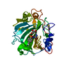
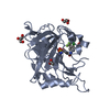
| Title | PHOSPHOLIPASE A2 INHIBITOR FROM VIPOXIN | ||||||
 Components Components | PHOSPHOLIPASE A2 INHIBITOR | ||||||
 Keywords Keywords | NEUROTOXIN / PHOSPHOLIPASE A2 INHIBITOR / RECOGNITION / MOLECULAR EVOLUTION | ||||||
| Function / homology |  Function and homology information Function and homology information: / arachidonate secretion / lipid catabolic process / negative regulation of T cell proliferation / phospholipid metabolic process / phospholipid binding / toxin activity / calcium ion binding / extracellular region Similarity search - Function | ||||||
| Biological species |  Vipera ammodytes (sand viper) Vipera ammodytes (sand viper) | ||||||
| Method |  X-RAY DIFFRACTION / X-RAY DIFFRACTION /  MOLECULAR REPLACEMENT / Resolution: 1.76 Å MOLECULAR REPLACEMENT / Resolution: 1.76 Å | ||||||
 Authors Authors | Devedjiev, Y.D. / Popov, A.N. | ||||||
 Citation Citation |  Journal: J.Mol.Biol. / Year: 1997 Journal: J.Mol.Biol. / Year: 1997Title: X-ray structure at 1.76 A resolution of a polypeptide phospholipase A2 inhibitor. Authors: Devedjiev, Y. / Popov, A. / Atanasov, B. / Bartunik, H.D. #1:  Journal: Eur.Cryst.Meeting / Year: 1994 Journal: Eur.Cryst.Meeting / Year: 1994Title: Structure and Mechanism of Phospholipase A2 Authors: Devedjiev, Y. / Popov, A. / Bartunik, H.-D. / Atanasov, B. #2:  Journal: J.Mol.Biol. / Year: 1993 Journal: J.Mol.Biol. / Year: 1993Title: Crystals of Phospholipase A2 Inhibitor. The Non-Toxic Component of Vipoxin from the Venom of Bulgarian Viper (Vipera Ammodytes) Authors: Devedjiev, Y. / Atanasov, B. / Mancheva, I. / Aleksiev, B. #3:  Journal: Dokl.Bolg.Akad.Nauk / Year: 1989 Journal: Dokl.Bolg.Akad.Nauk / Year: 1989Title: Solubility and Phase States of Vipoxin from the Venom of Bulgarian Viper (Vipera Ammodytes Ammodytes) Authors: Devedjiev, Y.D. / Mancheva, I.N. / Aleksiev, B.V. / Atanasov, B.P. | ||||||
| History |
|
- Structure visualization
| Structure viewer | Molecule:  Molmil Molmil Jmol/JSmol Jmol/JSmol |
|---|
- Downloads & links
- Download
| PDBx/mmCIF format |  1vpi.cif.gz 1vpi.cif.gz | 39.1 KB | Display |  PDBx/mmCIF format PDBx/mmCIF format |
|---|---|---|---|---|
| PDB format |  pdb1vpi.ent.gz pdb1vpi.ent.gz | 26.6 KB | Display |  PDB format PDB format |
| PDBx/mmJSON format |  1vpi.json.gz 1vpi.json.gz | Tree view |  PDBx/mmJSON format PDBx/mmJSON format | |
| Others |  Other downloads Other downloads |
-Validation report
| Arichive directory |  https://data.pdbj.org/pub/pdb/validation_reports/vp/1vpi https://data.pdbj.org/pub/pdb/validation_reports/vp/1vpi ftp://data.pdbj.org/pub/pdb/validation_reports/vp/1vpi ftp://data.pdbj.org/pub/pdb/validation_reports/vp/1vpi | HTTPS FTP |
|---|
-Related structure data
| Related structure data |  1pp2S S: Starting model for refinement |
|---|---|
| Similar structure data |
- Links
Links
- Assembly
| Deposited unit | 
| ||||||||||||
|---|---|---|---|---|---|---|---|---|---|---|---|---|---|
| 1 | 
| ||||||||||||
| Unit cell |
| ||||||||||||
| Components on special symmetry positions |
|
- Components
| #1: Protein | Mass: 13650.032 Da / Num. of mol.: 1 / Source method: isolated from a natural source / Source: (natural)  Vipera ammodytes (sand viper) / Organ: VENOM GLAND / References: UniProt: P04084 Vipera ammodytes (sand viper) / Organ: VENOM GLAND / References: UniProt: P04084 |
|---|---|
| #2: Water | ChemComp-HOH / |
| Has protein modification | Y |
-Experimental details
-Experiment
| Experiment | Method:  X-RAY DIFFRACTION / Number of used crystals: 1 X-RAY DIFFRACTION / Number of used crystals: 1 |
|---|
- Sample preparation
| Crystal | Density Matthews: 2.3 Å3/Da / Density % sol: 46 % | |||||||||||||||||||||||||
|---|---|---|---|---|---|---|---|---|---|---|---|---|---|---|---|---|---|---|---|---|---|---|---|---|---|---|
| Crystal grow | pH: 8.3 Details: PROTEIN WAS CRYSTALLIZED FROM 52% AMMONIUM SULFATE, 0.5% MPD, 100 MM TRIS, PH 8.3 | |||||||||||||||||||||||||
| Crystal grow | *PLUS Method: vapor diffusion, hanging drop | |||||||||||||||||||||||||
| Components of the solutions | *PLUS
|
-Data collection
| Diffraction | Mean temperature: 293 K |
|---|---|
| Diffraction source | Source: SEALED TUBE / Wavelength: 1.5418 |
| Detector | Type: OIIF, DUBNA, RUSSIA / Detector: AREA DETECTOR / Date: Jun 1, 1993 / Details: COLLIMATOR |
| Radiation | Monochromator: GRAPHITE(002) / Monochromatic (M) / Laue (L): M / Scattering type: x-ray |
| Radiation wavelength | Wavelength: 1.5418 Å / Relative weight: 1 |
| Reflection | Highest resolution: 1.76 Å / Num. obs: 11854 / % possible obs: 98 % / Observed criterion σ(I): 1 / Redundancy: 4.2 % / Biso Wilson estimate: 20.7 Å2 / Rmerge(I) obs: 0.062 / Rsym value: 0.058 |
| Reflection shell | Resolution: 1.76→1.86 Å / % possible all: 48 |
| Reflection | *PLUS Num. measured all: 42158 |
| Reflection shell | *PLUS % possible obs: 48 % |
- Processing
| Software |
| ||||||||||||||||||||||||||||||||||||||||||||||||||||||||||||
|---|---|---|---|---|---|---|---|---|---|---|---|---|---|---|---|---|---|---|---|---|---|---|---|---|---|---|---|---|---|---|---|---|---|---|---|---|---|---|---|---|---|---|---|---|---|---|---|---|---|---|---|---|---|---|---|---|---|---|---|---|---|
| Refinement | Method to determine structure:  MOLECULAR REPLACEMENT MOLECULAR REPLACEMENTStarting model: PDB ENTRY 1PP2 Resolution: 1.76→6 Å / σ(F): 2.5
| ||||||||||||||||||||||||||||||||||||||||||||||||||||||||||||
| Displacement parameters | Biso mean: 24.7 Å2 | ||||||||||||||||||||||||||||||||||||||||||||||||||||||||||||
| Refine analyze | Luzzati coordinate error obs: 0.18 Å / Luzzati d res low obs: 6 Å | ||||||||||||||||||||||||||||||||||||||||||||||||||||||||||||
| Refinement step | Cycle: LAST / Resolution: 1.76→6 Å
| ||||||||||||||||||||||||||||||||||||||||||||||||||||||||||||
| Refine LS restraints |
| ||||||||||||||||||||||||||||||||||||||||||||||||||||||||||||
| Software | *PLUS Name:  X-PLOR / Version: 3.1 / Classification: refinement X-PLOR / Version: 3.1 / Classification: refinement | ||||||||||||||||||||||||||||||||||||||||||||||||||||||||||||
| Refinement | *PLUS | ||||||||||||||||||||||||||||||||||||||||||||||||||||||||||||
| Solvent computation | *PLUS | ||||||||||||||||||||||||||||||||||||||||||||||||||||||||||||
| Displacement parameters | *PLUS | ||||||||||||||||||||||||||||||||||||||||||||||||||||||||||||
| Refine LS restraints | *PLUS
|
 Movie
Movie Controller


 PDBj
PDBj

