 Movie
Movie Controller

[English] 日本語
 Yorodumi
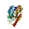
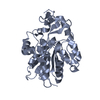
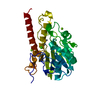
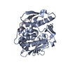
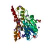
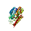
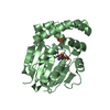
- PDB-1pp2: THE REFINED CRYSTAL STRUCTURE OF DIMERIC PHOSPHOLIPASE A2 AT 2.5 ... -
+ Open data

- Basic information
| Entry | Database: PDB / ID: 1pp2 | ||||||
|---|---|---|---|---|---|---|---|
| Title | THE REFINED CRYSTAL STRUCTURE OF DIMERIC PHOSPHOLIPASE A2 AT 2.5 ANGSTROMS. ACCESS TO A SHIELDED CATALYTIC CENTER | ||||||
 Components Components | CALCIUM-FREE PHOSPHOLIPASE A2 | ||||||
 Keywords Keywords | HYDROLASE | ||||||
| Function / homology |  Function and homology information Function and homology informationA2-type glycerophospholipase activity / phospholipase A2 / arachidonate secretion / lipid catabolic process / negative regulation of T cell proliferation / phospholipid metabolic process / phospholipid binding / toxin activity / calcium ion binding / extracellular region Similarity search - Function | ||||||
| Biological species |  Crotalus atrox (western diamondback rattlesnake) Crotalus atrox (western diamondback rattlesnake) | ||||||
| Method |  X-RAY DIFFRACTION / Resolution: 2.5 Å X-RAY DIFFRACTION / Resolution: 2.5 Å | ||||||
 Authors Authors | Brunie, S. / Sigler, P.B. | ||||||
 Citation Citation | Journal: J.Biol.Chem. / Year: 1985 Title: The refined crystal structure of dimeric phospholipase A2 at 2.5 A. Access to a shielded catalytic center. Authors: Brunie, S. / Bolin, J. / Gewirth, D. / Sigler, P.B. #1: Journal: J.Biol.Chem. / Year: 1985Title: A Comparison of the Crystal Structures of Phospholipase A2 from Bovine Pancreas and Crotalus Atrox Venom Authors: Renetseder, R. / Brunie, S. / Dijkstra, B.W. / Drenth, J. / Sigler, P.B. #2: Journal: J.Biol.Chem. / Year: 1981Title: The 2.5 Angstroms Crystal Structure of a Dimeric Phospholipase A2 from the Venom of Crotalus Atrox Authors: Keith, C. / Feldman, D.S. / Deganello, S. / Glick, J. / Ward, K.B. / Jones, E.O. / Sigler, P.B. #3: Journal: J.Mol.Biol. / Year: 1975Title: Characterization of Crystals of Two Venom Phospholipases A2 Authors: Pasek, M. / Keith, C. / Feldman, D. / Sigler, P.B. | ||||||
| History |
|
- Structure visualization
| Structure viewer | Molecule: MolmilJmol/JSmol |
|---|
- Downloads & links
-Download
| PDBx/mmCIF format | 1pp2.cif.gz | 63.1 KB | Display | PDBx/mmCIF format |
|---|---|---|---|---|
| PDB format | pdb1pp2.ent.gz | 45.4 KB | Display | PDB format |
| PDBx/mmJSON format | 1pp2.json.gz | Tree view | PDBx/mmJSON format | |
| Others |  Other downloads Other downloads |
-Validation report
| Arichive directory | https://data.pdbj.org/pub/pdb/validation_reports/pp/1pp2ftp://data.pdbj.org/pub/pdb/validation_reports/pp/1pp2 | HTTPS FTP |
|---|
-Related structure data
| Similar structure data |
|---|


-Links
 PDBj
PDBj

- Assembly
| Deposited unit | 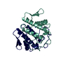
| ||||||||
|---|---|---|---|---|---|---|---|---|---|
| 1 |
| ||||||||
| Unit cell |
| ||||||||
| Noncrystallographic symmetry (NCS) | NCS oper: (Code: given Matrix: (-0.12725, 0.1029, -0.98652), Vector: Details | THE ENZYME IS A DIMER COMPOSED OF TWO IDENTICAL SUBUNITS OF 122 RESIDUES EACH. THE *RIGHT* SUBUNIT HAS BEEN ASSIGNED CHAIN IDENTIFIER *R*. THE *LEFT* SUBUNIT HAS BEEN ASSIGNED CHAIN IDENTIFIER *L*. THE TRANSFORMATION PRESENTED ON THE *MTRIX* RECORDS BELOW WILL YIELD APPROXIMATE COORDINATES FOR THE *RIGHT* SUBUNIT (CHAIN *R*) WHEN APPLIED TO THE *LEFT* SUBUNIT (CHAIN *L*). | |
-Components
| #1: Protein | Mass: 13607.277 Da / Num. of mol.: 2 Source method: isolated from a genetically manipulated source Source: (gene. exp.) Crotalus atrox (western diamondback rattlesnake)References: UniProt: P00624, phospholipase A2 #2: Water | ChemComp-HOH / |  Mass: 18.015 Da / Num. of mol.: 137 / Source method: isolated from a natural source / Formula: H2O Mass: 18.015 Da / Num. of mol.: 137 / Source method: isolated from a natural source / Formula: H2OHas protein modification | Y | |
|---|
-Experimental details
-Experiment
| Experiment | Method: X-RAY DIFFRACTION |
|---|
- Sample preparation
| Crystal | Density Matthews: 2.39 Å3/Da / Density % sol: 48.48 % | ||||||||||||||||||||||||||||||||||||||||||
|---|---|---|---|---|---|---|---|---|---|---|---|---|---|---|---|---|---|---|---|---|---|---|---|---|---|---|---|---|---|---|---|---|---|---|---|---|---|---|---|---|---|---|---|
| Crystal grow | *PLUS Temperature: 4 ℃ / pH: 7.2 / Method: microdialysis | ||||||||||||||||||||||||||||||||||||||||||
| Components of the solutions | *PLUS
|
-Data collection
| Radiation | Scattering type: x-ray |
|---|---|
| Radiation wavelength | Relative weight: 1 |
- Processing
| Software | Name: PROLSQ / Classification: refinement | |||||||||||||||||||||||||||||||||||||||||||||||||||||||||||||||
|---|---|---|---|---|---|---|---|---|---|---|---|---|---|---|---|---|---|---|---|---|---|---|---|---|---|---|---|---|---|---|---|---|---|---|---|---|---|---|---|---|---|---|---|---|---|---|---|---|---|---|---|---|---|---|---|---|---|---|---|---|---|---|---|---|
| Refinement | Rfactor obs: 0.178 / Highest resolution: 2.5 Å | |||||||||||||||||||||||||||||||||||||||||||||||||||||||||||||||
| Refinement step | Cycle: LAST / Highest resolution: 2.5 Å
| |||||||||||||||||||||||||||||||||||||||||||||||||||||||||||||||
| Refine LS restraints |
| |||||||||||||||||||||||||||||||||||||||||||||||||||||||||||||||
| Refinement | *PLUS σ(I): 2 | |||||||||||||||||||||||||||||||||||||||||||||||||||||||||||||||
| Solvent computation | *PLUS | |||||||||||||||||||||||||||||||||||||||||||||||||||||||||||||||
| Displacement parameters | *PLUS |
