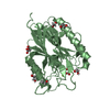
 Movie
Movie Controller


+ Open data

- Basic information
| Entry | Database: PDB / ID: 1jlt | ||||||
|---|---|---|---|---|---|---|---|
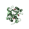
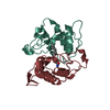
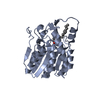
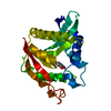
| Title | Vipoxin Complex | ||||||
 Components Components |
| ||||||
 Keywords Keywords | HYDROLASE / HETERODIMER COMPLEX / PHOSPHOLIPASE / VIPOXIN / PLA2-ACTIVITY | ||||||
| Function / homology |  Function and homology information Function and homology information: / phospholipase A2 / arachidonate secretion / lipid catabolic process / negative regulation of T cell proliferation / phospholipid metabolic process / phospholipid binding / toxin activity / killing of cells of another organism / calcium ion binding / extracellular region Similarity search - Function | ||||||
| Biological species |  Vipera ammodytes ammodytes (western sand viper) Vipera ammodytes ammodytes (western sand viper) | ||||||
| Method |  X-RAY DIFFRACTION / SYNCHROTRON / MOLECULAR REPLACEMENT / Resolution: 1.4 Å X-RAY DIFFRACTION / SYNCHROTRON / MOLECULAR REPLACEMENT / Resolution: 1.4 Å | ||||||
 Authors Authors | Banumathi, S. / Rajashankar, K.R. / Notzel, C. / Aleksiev, B. / Singh, T.P. / Genov, N. / Betzel, C. | ||||||
 Citation Citation | Journal: Acta Crystallogr.,Sect.D / Year: 2001 Title: Structure of the neurotoxic complex vipoxin at 1.4 A resolution. Authors: Banumathi, S. / Rajashankar, K.R. / Notzel, C. / Aleksiev, B. / Singh, T.P. / Genov, N. / Betzel, C. | ||||||
| History |
| ||||||
| Remark 999 | sequence The authors' residue numbering is not sequential. Residue numbers 15, 57, 60, 62-67, 87, ...sequence The authors' residue numbering is not sequential. Residue numbers 15, 57, 60, 62-67, 87, 123 are not used in the coordinates. |
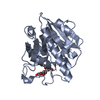
- Structure visualization
| Structure viewer | Molecule: MolmilJmol/JSmol |
|---|
- Downloads & links
-Download
| PDBx/mmCIF format | 1jlt.cif.gz | 68.9 KB | Display | PDBx/mmCIF format |
|---|---|---|---|---|
| PDB format | pdb1jlt.ent.gz | 49.7 KB | Display | PDB format |
| PDBx/mmJSON format | 1jlt.json.gz | Tree view | PDBx/mmJSON format | |
| Others |  Other downloads Other downloads |
-Validation report
| Arichive directory | https://data.pdbj.org/pub/pdb/validation_reports/jl/1jltftp://data.pdbj.org/pub/pdb/validation_reports/jl/1jlt | HTTPS FTP |
|---|
-Related structure data
| Related structure data |  1aokS S: Starting model for refinement |
|---|---|
| Similar structure data |


-Links
 PDBj
PDBj

- Assembly
| Deposited unit | 
| ||||||||
|---|---|---|---|---|---|---|---|---|---|
| 1 |
| ||||||||
| Unit cell |
|
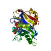
-Components
| #1: Protein | Mass: 13664.058 Da / Num. of mol.: 1 / Source method: isolated from a natural source Source: (natural) Vipera ammodytes ammodytes (western sand viper)Species: Vipera ammodytes / Strain: ammodytes / References: UniProt: P04084 |
|---|---|
| #2: Protein | Mass: 13846.822 Da / Num. of mol.: 1 / Source method: isolated from a natural source Details: Sequence homology between phospholipase and its inhibitor in snake venom. The primary structure of phospholipase A2 of vipoxin from the venom of the Bulgarian viper (Vipera ammodytes ammodytes, Serpentes) Source: (natural) Vipera ammodytes ammodytes (western sand viper)Species: Vipera ammodytes / Strain: ammodytes / References: UniProt: P14420, phospholipase A2 |
| #3: Chemical | ChemComp-MRD / (  Mass: 118.174 Da / Num. of mol.: 1 / Source method: obtained synthetically / Formula: C6H14O2 / Comment: precipitant*YM Mass: 118.174 Da / Num. of mol.: 1 / Source method: obtained synthetically / Formula: C6H14O2 / Comment: precipitant*YM |
| #4: Chemical | ChemComp-MPD / (  Mass: 118.174 Da / Num. of mol.: 1 / Source method: obtained synthetically / Formula: C6H14O2 / Comment: precipitant*YM Mass: 118.174 Da / Num. of mol.: 1 / Source method: obtained synthetically / Formula: C6H14O2 / Comment: precipitant*YM |
| #5: Water | ChemComp-HOH /  Mass: 18.015 Da / Num. of mol.: 320 / Source method: isolated from a natural source / Formula: H2O Mass: 18.015 Da / Num. of mol.: 320 / Source method: isolated from a natural source / Formula: H2O |
| Has protein modification | Y |
-Experimental details
-Experiment
| Experiment | Method: X-RAY DIFFRACTION / Number of used crystals: 3 |
|---|
- Sample preparation
| Crystal | Density Matthews: 2.32 Å3/Da / Density % sol: 47 % | ||||||||||||||||||||
|---|---|---|---|---|---|---|---|---|---|---|---|---|---|---|---|---|---|---|---|---|---|
| Crystal grow | Temperature: 277 K / Method: vapor diffusion, hanging drop / pH: 4.8 Details: PEG 4000, MPD, pH 4.8, VAPOR DIFFUSION, HANGING DROP | ||||||||||||||||||||
| Crystal grow | *PLUS Temperature: 287 K | ||||||||||||||||||||
| Components of the solutions | *PLUS
|
-Data collection
| Diffraction | Mean temperature: 100 K |
|---|---|
| Diffraction source | Source: SYNCHROTRON / Site: EMBL/DESY, HAMBURG  / Beamline: X11 / Wavelength: 0.908 Å / Beamline: X11 / Wavelength: 0.908 Å |
| Detector | Type: MARRESEARCH / Detector: IMAGE PLATE / Date: Jan 1, 2000 / Details: Collimator |
| Radiation | Protocol: SINGLE WAVELENGTH / Monochromatic (M) / Laue (L): M / Scattering type: x-ray |
| Radiation wavelength | Wavelength: 0.908 Å / Relative weight: 1 |
| Reflection | Resolution: 1.37→20 Å / Num. all: 54019 / Num. obs: 53423 / % possible obs: 99.1 % / Redundancy: 4.3 % / Biso Wilson estimate: 12.7 Å2 / Rmerge(I) obs: 0.067 / Net I/σ(I): 23 |
| Reflection shell | Resolution: 1.37→1.39 Å / Redundancy: 3 % / Rmerge(I) obs: 0.257 / Mean I/σ(I) obs: 4 / Num. unique all: 2525 / % possible all: 96.1 |
| Reflection | *PLUS Lowest resolution: 20 Å |
| Reflection shell | *PLUS % possible obs: 96.1 % |
- Processing
| Software |
| ||||||||||||||||||||
|---|---|---|---|---|---|---|---|---|---|---|---|---|---|---|---|---|---|---|---|---|---|
| Refinement | Method to determine structure: MOLECULAR REPLACEMENT Starting model: PDB ENTRY 1AOK Resolution: 1.4→20 Å / Isotropic thermal model: Isotropic / Cross valid method: THROUGHOUT Details: used maximum likelihood target using amplitudes procedure
| ||||||||||||||||||||
| Displacement parameters | Biso mean: 15.7 Å2
| ||||||||||||||||||||
| Refine analyze | Luzzati coordinate error obs: 0.15 Å / Luzzati d res low obs: 5 Å / Luzzati sigma a obs: 0.08 Å | ||||||||||||||||||||
| Refinement step | Cycle: LAST / Resolution: 1.4→20 Å
| ||||||||||||||||||||
| Refine LS restraints |
| ||||||||||||||||||||
| Software | *PLUS Name: CNS / Classification: refinement | ||||||||||||||||||||
| Refinement | *PLUS Highest resolution: 1.4 Å / Lowest resolution: 20 Å / % reflection Rfree: 10 % / Rfactor obs: 0.182 | ||||||||||||||||||||
| Solvent computation | *PLUS | ||||||||||||||||||||
| Displacement parameters | *PLUS Biso mean: 15.7 Å2 | ||||||||||||||||||||
| Refine LS restraints | *PLUS
|
