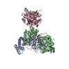
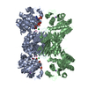
| Entry | Database: PDB / ID: 1vm6
|
|---|
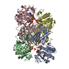
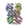
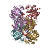
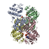
| Title | Crystal structure of Dihydrodipicolinate reductase (TM1520) from Thermotoga maritima at 2.27 A resolution |
|---|
 Components Components | Dihydrodipicolinate reductase |
|---|
 Keywords Keywords | OXIDOREDUCTASE / TM1520 / DIHYDRODIPICOLINATE REDUCTASE / STRUCTURAL GENOMICS / JCSG / PROTEIN STRUCTURE INITIATIVE / PSI / Joint Center for Structural Genomics |
|---|
| Function / homology |  Function and homology information Function and homology information
4-hydroxy-tetrahydrodipicolinate reductase / oxidoreductase activity, acting on CH or CH2 groups, NAD or NADP as acceptor / 4-hydroxy-tetrahydrodipicolinate reductase activity / : / : / NAD binding / NADP binding / cytosolSimilarity search - Function Dihydrodipicolinate reductase, conserved site / Dihydrodipicolinate reductase signature. / Dihydrodipicolinate reductase, C-terminal / Dihydrodipicolinate reductase / Dihydrodipicolinate reductase, C-terminus / Dihydrodipicolinate reductase, N-terminal / Dihydrodipicolinate reductase, N-terminus / Dihydrodipicolinate Reductase; domain 2 / Dihydrodipicolinate Reductase; domain 2 / NAD(P)-binding Rossmann-like Domain ...Dihydrodipicolinate reductase, conserved site / Dihydrodipicolinate reductase signature. / Dihydrodipicolinate reductase, C-terminal / Dihydrodipicolinate reductase / Dihydrodipicolinate reductase, C-terminus / Dihydrodipicolinate reductase, N-terminal / Dihydrodipicolinate reductase, N-terminus / Dihydrodipicolinate Reductase; domain 2 / Dihydrodipicolinate Reductase; domain 2 / NAD(P)-binding Rossmann-like Domain / NAD(P)-binding domain superfamily / Rossmann fold / 2-Layer Sandwich / 3-Layer(aba) Sandwich / Alpha BetaSimilarity search - Domain/homology |
|---|
| Biological species |   Thermotoga maritima (bacteria) Thermotoga maritima (bacteria) |
|---|
| Method |  X-RAY DIFFRACTION / SYNCHROTRON / MOLECULAR REPLACEMENT / Resolution: 2.27 Å X-RAY DIFFRACTION / SYNCHROTRON / MOLECULAR REPLACEMENT / Resolution: 2.27 Å |
|---|
 Authors Authors | Joint Center for Structural Genomics (JCSG) |
|---|
 Citation Citation | Journal: To be published
Title: Crystal structure of Dihydrodipicolinate reductase (TM1520) from Thermotoga maritima at 2.27 A resolution
Authors: Joint Center for Structural Genomics (JCSG) |
|---|
| History | | Deposition | Sep 3, 2004 | Deposition site: RCSB / Processing site: RCSB |
|---|
| Revision 1.0 | Sep 21, 2004 | Provider: repository / Type: Initial release |
|---|
| Revision 1.1 | Apr 26, 2008 | Group: Version format compliance |
|---|
| Revision 1.2 | Jul 13, 2011 | Group: Advisory / Version format compliance |
|---|
| Revision 1.3 | Oct 4, 2017 | Group: Refinement description / Category: software
Item: _software.classification / _software.name / _software.version |
|---|
| Revision 1.4 | Jan 25, 2023 | Group: Database references / Derived calculations / Category: database_2 / struct_ref_seq_dif / struct_site
Item: _database_2.pdbx_DOI / _database_2.pdbx_database_accession ..._database_2.pdbx_DOI / _database_2.pdbx_database_accession / _struct_ref_seq_dif.details / _struct_site.pdbx_auth_asym_id / _struct_site.pdbx_auth_comp_id / _struct_site.pdbx_auth_seq_id |
|---|
| Revision 1.5 | Sep 20, 2023 | Group: Data collection / Refinement description
Category: chem_comp_atom / chem_comp_bond ...chem_comp_atom / chem_comp_bond / pdbx_initial_refinement_model / struct_ncs_dom_lim
Item: _struct_ncs_dom_lim.beg_auth_comp_id / _struct_ncs_dom_lim.end_auth_comp_id |
|---|
|
|---|
 Movie
Movie Controller

 Yorodumi
 Open data
 Basic information
 Structure visualization
 Downloads & links
 Other downloads


 PDBj
PDBj Assembly


 Mass: 59.044 Da / Num. of mol.: 12 / Source method: obtained synthetically / Formula: C2H3O2
 Mass: 663.425 Da / Num. of mol.: 4 / Source method: obtained synthetically / Formula: C21H27N7O14P2 / Comment: NAD*YM
 Mass: 194.226 Da / Num. of mol.: 4 / Source method: obtained synthetically / Formula: C8H18O5 / Comment: precipitant*YM
 Mass: 62.068 Da / Num. of mol.: 1 / Source method: obtained synthetically / Formula: C2H6O2
 Sample preparation
 / Beamline: BL11-1 / Wavelength: 0.980075
  Processing