Resolution: 2.1→29.56 Å / Num. obs: 293140 / % possible obs: 93 % / Redundancy: 3.8 % / Biso Wilson estimate: 32.49 Å2 / Rsym value: 0.09 / Net I/σ(I): 9.1
Reflection shell
Resolution: 2.1→2.21 Å / Redundancy: 3.5 % / Mean I/σ(I) obs: 2.6 / Num. unique all: 30908 / Rsym value: 0.433 / % possible all: 67.2
-
Processing
Software
Name
Version
Classification
MOSFLM
datareduction
SCALA
4.2)
datascaling
SHARP
phasing
autoSHARP
phasing
SHELX
modelbuilding
WARP
modelbuilding
REFMAC
5.2.0005
refinement
CCP4
(SCALA)
datascaling
SHELX
phasing
ARP/wARP
modelbuilding
Refinement
Method to determine structure: MAD / Resolution: 2.1→29.56 Å / Cor.coef. Fo:Fc: 0.967 / Cor.coef. Fo:Fc free: 0.947 / SU B: 5.076 / SU ML: 0.128 / Cross valid method: THROUGHOUT / ESU R: 0.195 / ESU R Free: 0.17 Stereochemistry target values: MAXIMUM LIKELIHOOD WITH PHASES Details: 1. HYDROGENS HAVE BEEN ADDED IN THE RIDING POSITIONS 2. THE ACTIVE SITE TRIAD CONSISTS OF SER188, HIS303 AND ASP274. THE ELECTRON DENSITY SUGGESTED THERE COULD EXIST A PARTIALLY OCCUPIED ...Details: 1. HYDROGENS HAVE BEEN ADDED IN THE RIDING POSITIONS 2. THE ACTIVE SITE TRIAD CONSISTS OF SER188, HIS303 AND ASP274. THE ELECTRON DENSITY SUGGESTED THERE COULD EXIST A PARTIALLY OCCUPIED ACYL INTERMEDIATE ON SER188. HOWEVER, IT IS NOT CONCLUSIVE. AS A RESULT, WATER MOLECULES WERE MODELLED. 3. THERE IS SOME UNEXPLAINED DENSITY NEAR N-TERMINUS 4. RESIDUES 134,135 OF CHAIN D,H,I,J,K,L HAVE POOR DENSITY
Rfactor
Num. reflection
% reflection
Selection details
Rfree
0.22336
14726
5 %
RANDOM
Rwork
0.18354
-
-
-
obs
0.18554
278371
92.77 %
-
Solvent computation
Ion probe radii: 0.8 Å / Shrinkage radii: 0.8 Å / VDW probe radii: 1.2 Å / Solvent model: BABINET MODEL WITH MASK
 Movie
Movie Controller

 Yorodumi
 Open data
 Basic information
 Components
 Keywords
 Function and homology information
 Thermotoga maritima (bacteria)
 X-RAY DIFFRACTION /
  Authors
 Citation
 Structure visualization
 Downloads & links
 Other downloads


 PDBj
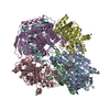
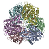
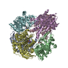
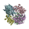
PDBj Assembly
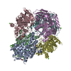
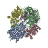
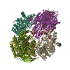
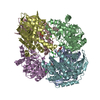
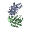
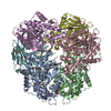
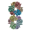

 Mass: 92.094 Da / Num. of mol.: 1 / Source method: obtained synthetically / Formula: C3H8O3
 Mass: 18.015 Da / Num. of mol.: 2464 / Source method: isolated from a natural source / Formula: H2O
 Sample preparation
 Processing