 Movie
Movie Controller


+ Open data

- Basic information
| Entry | Database: PDB / ID: 1vkl | ||||||
|---|---|---|---|---|---|---|---|
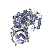
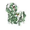
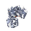
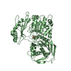
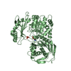
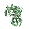
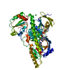
| Title | RABBIT MUSCLE PHOSPHOGLUCOMUTASE | ||||||
 Components Components | PHOSPHOGLUCOMUTASE | ||||||
 Keywords Keywords | PHOSPHOTRANSFERASE / PHOSPHOGLUCOMUTASE | ||||||
| Function / homology |  Function and homology information Function and homology informationphosphoglucomutase (alpha-D-glucose-1,6-bisphosphate-dependent) / phosphoglucomutase activity / sarcoplasmic reticulum / glucose metabolic process / magnesium ion binding / cytosol Similarity search - Function | ||||||
| Biological species |  | ||||||
| Method |  X-RAY DIFFRACTION / Resolution: 2.7 Å X-RAY DIFFRACTION / Resolution: 2.7 Å | ||||||
 Authors Authors | Ray Junior, W.J. / Baranidharan, S. / Liu, Y. | ||||||
 Citation Citation | Journal: Biochemistry / Year: 1993 Title: Structural changes at the metal ion binding site during the phosphoglucomutase reaction. Authors: Ray Jr., W.J. / Post, C.B. / Liu, Y. / Rhyu, G.I. #1: Journal: J.Biol.Chem. / Year: 1992Title: The Crystal Structure of Muscle Phosphoglucomutase Refined at 2.7-Angstrom Resolution Authors: Dai, J.B. / Liu, Y. / Ray Junior, W.J. / Konno, M. #2: Journal: J.Biol.Chem. / Year: 1986Title: The Catalytic Activity of Muscle Phosphoglucomutase in the Crystalline Phase Authors: Ray Junior, W.J. #3: Journal: J.Biol.Chem. / Year: 1986Title: The Structure of Rabbit Muscle Phosphoglucomutase at Intermediate Resolution Authors: Lin, Z. / Konno, M. / Abad-Zapatero, C. / Wierenga, R. / Murthy, M.R. / Ray Junior, W.J. / Rossmann, M.G. | ||||||
| History |
| ||||||
| Remark 650 | HELIX THE MONOMER CAN BE SUBDIVIDED INTO FOUR SEQUENCE DOMAINS: THE FINAL COLUMN OF THE HELIX ...HELIX THE MONOMER CAN BE SUBDIVIDED INTO FOUR SEQUENCE DOMAINS: THE FINAL COLUMN OF THE HELIX IDENTIFIER, 1-4, DESIGNATES THE DOMAINS; FOR STRANDS, THE DOMAINS ARE DESIGNATED BY THE NUMBERS IN THE SECOND COLUMN, 1-4, FOLLOWED BY EITHER B OR A SPATIAL RELATIONSHIP EXISTS BETWEEN GROUPS OF HELICES/STR IN DOMAINS 1-3. IN ORDER TO EMPHASIZE THIS RELATIONSHIP, AS 4S-1S. THUS, A SPATIAL DOMAIN-DOMAIN RELATIONSHIP EXIST AMONG STANDS/HELICES WHOSE DESIGNATOR CONTAINS 1S-4S IN THE SECOND COLUMN OR ENDS WITH 1-4, RESPECTIVELY. DOMAINS 1, 2, 3 IN MONOMER 1 AND MONOMER 2 ARE RELATED BY A ROTATION MATRIX GIVEN AS MTRIX1. DOMAIN 4 IN MONOMER 1 AND MONOMER 2 ARE RELATED BY A DIFFERENT ROTATION MATRIX GIVEN AS MTRIX2. |
- Structure visualization
| Structure viewer | Molecule: MolmilJmol/JSmol |
|---|
- Downloads & links
-Download
| PDBx/mmCIF format | 1vkl.cif.gz | 231.3 KB | Display | PDBx/mmCIF format |
|---|---|---|---|---|
| PDB format | pdb1vkl.ent.gz | 185.4 KB | Display | PDB format |
| PDBx/mmJSON format | 1vkl.json.gz | Tree view | PDBx/mmJSON format | |
| Others |  Other downloads Other downloads |
-Validation report
| Arichive directory | https://data.pdbj.org/pub/pdb/validation_reports/vk/1vklftp://data.pdbj.org/pub/pdb/validation_reports/vk/1vkl | HTTPS FTP |
|---|
-Related structure data
| Related structure data | 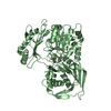 3pmgS S: Starting model for refinement |
|---|---|
| Similar structure data |


-Links
 PDBj
PDBj- Assembly
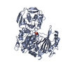
| Deposited unit | 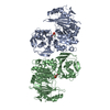
| ||||||||||||
|---|---|---|---|---|---|---|---|---|---|---|---|---|---|
| 1 | 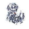
| ||||||||||||
| 2 | 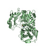
| ||||||||||||
| Unit cell |
| ||||||||||||
| Noncrystallographic symmetry (NCS) | NCS oper:
| ||||||||||||
| Details | THERE ARE TWO MONOMER COPIES PER ASYMMETRIC UNIT ALONG THE 4(1) SCREW AXIS. MONOMER A IS THE FIRST ENCOUNTERED IN AN ASYMMETRIC UNIT AS ONE MOVES CLOCKWISE ALONG A SCREW AXIS. AMINO ACID RESIDUES IN MONOMERS A AND B ARE DISTINGUISHED BY THE CHAIN IDENTIFIERS *A* AND *B*, RESPECTIVELY. |
-Components
| #1: Protein | Mass: 61579.902 Da / Num. of mol.: 2 / Source method: isolated from a natural source / Source: (natural) References: UniProt: P00949, phosphoglucomutase (alpha-D-glucose-1,6-bisphosphate-dependent) #2: Chemical |   Mass: 58.693 Da / Num. of mol.: 2 / Source method: obtained synthetically / Formula: Ni Mass: 58.693 Da / Num. of mol.: 2 / Source method: obtained synthetically / Formula: Ni#3: Water | ChemComp-HOH / |  Mass: 18.015 Da / Num. of mol.: 351 / Source method: isolated from a natural source / Formula: H2O Mass: 18.015 Da / Num. of mol.: 351 / Source method: isolated from a natural source / Formula: H2OHas protein modification | Y | |
|---|
-Experimental details
-Experiment
| Experiment | Method: X-RAY DIFFRACTION |
|---|
- Sample preparation
| Crystal | Density Matthews: 3.12 Å3/Da / Density % sol: 61 % | |||||||||||||||
|---|---|---|---|---|---|---|---|---|---|---|---|---|---|---|---|---|
| Crystal grow | pH: 6.4 / Details: pH 6.4 | |||||||||||||||
| Crystal | *PLUS | |||||||||||||||
| Crystal grow | *PLUS Method: unknown / Details: Lin, Z., (1986) J.Biol.Chem., 261, 264. | |||||||||||||||
| Components of the solutions | *PLUS
|
-Data collection
| Diffraction | Mean temperature: 289 K |
|---|---|
| Diffraction source | Wavelength: 1.5418 |
| Detector | Type: XUONG-HAMLIN MULTIWIRE / Detector: AREA DETECTOR / Date: Oct 1, 1993 |
| Radiation | Monochromatic (M) / Laue (L): M / Scattering type: x-ray |
| Radiation wavelength | Wavelength: 1.5418 Å / Relative weight: 1 |
| Reflection | Resolution: 2.6→20 Å / Num. obs: 46280 / % possible obs: 97 % / Observed criterion σ(I): 1 / Rmerge(I) obs: 0.15 |
- Processing
| Software |
| ||||||||||||||||||||||||||||||||||||||||||||||||||||||||||||
|---|---|---|---|---|---|---|---|---|---|---|---|---|---|---|---|---|---|---|---|---|---|---|---|---|---|---|---|---|---|---|---|---|---|---|---|---|---|---|---|---|---|---|---|---|---|---|---|---|---|---|---|---|---|---|---|---|---|---|---|---|---|
| Refinement | Starting model: PDB ENTRY 3PMG Resolution: 2.7→6 Å / σ(F): 2 Details: MOSTLY X-PLOR DEFAULT VALUES THE MODEL CONTAINS TEN RESIDUES, OUT OF 1122, THAT FALL IN THE GENEROUSLY ALLOWED REGION OF A RAMACHANDRAN PLOT AS DEFINED IN PROCHECK AND TWO RESIDUES IN THE ...Details: MOSTLY X-PLOR DEFAULT VALUES THE MODEL CONTAINS TEN RESIDUES, OUT OF 1122, THAT FALL IN THE GENEROUSLY ALLOWED REGION OF A RAMACHANDRAN PLOT AS DEFINED IN PROCHECK AND TWO RESIDUES IN THE DISALLOWED REGION. THE TWO RESIDUES IN THE DISALLOWED REGION ARE SEP B 116 AND ASN B 461.
| ||||||||||||||||||||||||||||||||||||||||||||||||||||||||||||
| Displacement parameters | Biso mean: 34 Å2 | ||||||||||||||||||||||||||||||||||||||||||||||||||||||||||||
| Refinement step | Cycle: LAST / Resolution: 2.7→6 Å
| ||||||||||||||||||||||||||||||||||||||||||||||||||||||||||||
| Refine LS restraints |
| ||||||||||||||||||||||||||||||||||||||||||||||||||||||||||||
| Xplor file |
| ||||||||||||||||||||||||||||||||||||||||||||||||||||||||||||
| Software | *PLUS Name: X-PLOR / Classification: refinement | ||||||||||||||||||||||||||||||||||||||||||||||||||||||||||||
| Refinement | *PLUS Rfactor obs: 0.17 / Rfactor Rfree: 0.21 / Rfactor Rwork: 0.17 | ||||||||||||||||||||||||||||||||||||||||||||||||||||||||||||
| Solvent computation | *PLUS | ||||||||||||||||||||||||||||||||||||||||||||||||||||||||||||
| Displacement parameters | *PLUS | ||||||||||||||||||||||||||||||||||||||||||||||||||||||||||||
| Refine LS restraints | *PLUS
|
