 Movie
Movie Controller


+ Open data

- Basic information
| Entry | Database: PDB / ID: 1v8i | ||||||
|---|---|---|---|---|---|---|---|
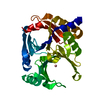
| Title | Crystal Structure Analysis of the ADP-ribose pyrophosphatase | ||||||
 Components Components | ADP-ribose pyrophosphatase | ||||||
 Keywords Keywords | HYDROLASE / Nudix motif / bacteria / RIKEN Structural Genomics/Proteomics Initiative / RSGI / Structural Genomics | ||||||
| Function / homology |  Function and homology information Function and homology informationpyrophosphatase activity / nucleoside phosphate metabolic process / ribose phosphate metabolic process / nucleotide binding / metal ion binding / cytosol Similarity search - Function | ||||||
| Biological species |   Thermus thermophilus (bacteria) Thermus thermophilus (bacteria) | ||||||
| Method |  X-RAY DIFFRACTION / SYNCHROTRON / MAD / Resolution: 1.76 Å X-RAY DIFFRACTION / SYNCHROTRON / MAD / Resolution: 1.76 Å | ||||||
 Authors Authors | Yoshiba, S. / Ooga, T. / Nakagawa, N. / Shibata, T. / Inoue, Y. / Yokoyama, S. / Kuramitsu, S. / Masui, R. / RIKEN Structural Genomics/Proteomics Initiative (RSGI) | ||||||
 Citation Citation | Journal: J.Biol.Chem. / Year: 2004 Title: Structural insights into the Thermus thermophilus ADP-ribose pyrophosphatase mechanism via crystal structures with the bound substrate and metal Authors: Yoshiba, S. / Ooga, T. / Nakagawa, N. / Shibata, T. / Inoue, Y. / Yokoyama, S. / Kuramitsu, S. / Masui, R. | ||||||
| History |
|
- Structure visualization
| Structure viewer | Molecule: MolmilJmol/JSmol |
|---|
- Downloads & links
-Download
| PDBx/mmCIF format | 1v8i.cif.gz | 42.9 KB | Display | PDBx/mmCIF format |
|---|---|---|---|---|
| PDB format | pdb1v8i.ent.gz | 30.2 KB | Display | PDB format |
| PDBx/mmJSON format | 1v8i.json.gz | Tree view | PDBx/mmJSON format | |
| Others |  Other downloads Other downloads |
-Validation report
| Summary document | 1v8i_validation.pdf.gz | 421.2 KB | Display | wwPDB validaton report |
|---|---|---|---|---|
| Full document | 1v8i_full_validation.pdf.gz | 422.6 KB | Display | |
| Data in XML | 1v8i_validation.xml.gz | 8 KB | Display | |
| Data in CIF | 1v8i_validation.cif.gz | 10.3 KB | Display | |
| Arichive directory | https://data.pdbj.org/pub/pdb/validation_reports/v8/1v8iftp://data.pdbj.org/pub/pdb/validation_reports/v8/1v8i | HTTPS FTP |
-Related structure data
| Related structure data | 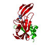 1v8lC 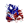 1v8mC 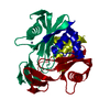 1v8nC 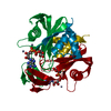 1v8rC 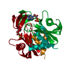 1v8sC  1v8tC 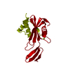 1v8uC 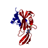 1v8vC 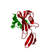 1v8wC  1v8yC C: citing same article ( |
|---|---|
| Similar structure data | |
| Other databases |


-Links
 PDBj
PDBj

- Assembly
| Deposited unit | 
| ||||||||
|---|---|---|---|---|---|---|---|---|---|
| 1 | 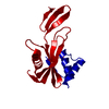
| ||||||||
| Unit cell |
|
-Components
| #1: Protein | Mass: 19287.912 Da / Num. of mol.: 1 Source method: isolated from a genetically manipulated source Source: (gene. exp.) Thermus thermophilus (bacteria) / Plasmid: pET11a / Species (production host): Escherichia coli / Production host: |
|---|---|
| #2: Water | ChemComp-HOH /  Mass: 18.015 Da / Num. of mol.: 59 / Source method: isolated from a natural source / Formula: H2O Mass: 18.015 Da / Num. of mol.: 59 / Source method: isolated from a natural source / Formula: H2O |
-Experimental details
-Experiment
| Experiment | Method: X-RAY DIFFRACTION / Number of used crystals: 1 |
|---|
- Sample preparation
| Crystal | Density Matthews: 2.36 Å3/Da / Density % sol: 47.55 % |
|---|---|
| Crystal grow | Temperature: 293 K / Method: vapor diffusion, hanging drop / pH: 5 Details: PEG 4000, ammonium sulfate, pH 5.0, VAPOR DIFFUSION, HANGING DROP, temperature 293K |
-Data collection
| Diffraction | Mean temperature: 103 K | |||||||||||||||
|---|---|---|---|---|---|---|---|---|---|---|---|---|---|---|---|---|
| Diffraction source | Source: SYNCHROTRON / Site: SPring-8  / Beamline: BL44B2 / Wavelength: 0.9804, 0.9808, 0.9763, 0.9822 / Beamline: BL44B2 / Wavelength: 0.9804, 0.9808, 0.9763, 0.9822 | |||||||||||||||
| Detector | Type: MARRESEARCH / Detector: CCD | |||||||||||||||
| Radiation | Protocol: MAD / Monochromatic (M) / Laue (L): M / Scattering type: x-ray | |||||||||||||||
| Radiation wavelength |
| |||||||||||||||
| Reflection | Resolution: 1.76→50 Å / Num. obs: 17390 / Observed criterion σ(I): -3 / Biso Wilson estimate: 11.9 Å2 / Rsym value: 0.03 | |||||||||||||||
| Reflection shell | Resolution: 1.76→1.82 Å / Rsym value: 0.088 |
- Processing
| Software |
| ||||||||||||||||||||||||||||||||||||
|---|---|---|---|---|---|---|---|---|---|---|---|---|---|---|---|---|---|---|---|---|---|---|---|---|---|---|---|---|---|---|---|---|---|---|---|---|---|
| Refinement | Method to determine structure: MAD / Resolution: 1.76→29 Å / Rfactor Rfree error: 0.006 / Data cutoff high absF: 1401759.64 / Data cutoff low absF: 0 / Isotropic thermal model: RESTRAINED / Cross valid method: THROUGHOUT / σ(F): 0
| ||||||||||||||||||||||||||||||||||||
| Solvent computation | Solvent model: FLAT MODEL / Bsol: 50.3534 Å2 / ksol: 0.40639 e/Å3 | ||||||||||||||||||||||||||||||||||||
| Displacement parameters | Biso mean: 17.6 Å2
| ||||||||||||||||||||||||||||||||||||
| Refine analyze |
| ||||||||||||||||||||||||||||||||||||
| Refinement step | Cycle: LAST / Resolution: 1.76→29 Å
| ||||||||||||||||||||||||||||||||||||
| Refine LS restraints |
| ||||||||||||||||||||||||||||||||||||
| LS refinement shell | Resolution: 1.76→1.87 Å / Rfactor Rfree error: 0.015 / Total num. of bins used: 6
| ||||||||||||||||||||||||||||||||||||
| Xplor file |
|
