 Movie
Movie Controller

[English] 日本語
 Yorodumi
- PDB-1v8l: Structure Analysis of the ADP-ribose pyrophosphatase complexed wi... -
+ Open data

- Basic information
| Entry | Database: PDB / ID: 1v8l | ||||||
|---|---|---|---|---|---|---|---|
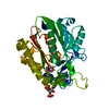
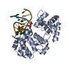
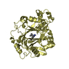
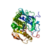
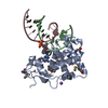
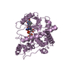
| Title | Structure Analysis of the ADP-ribose pyrophosphatase complexed with ADP-ribose | ||||||
 Components Components | ADP-ribose pyrophosphatase | ||||||
 Keywords Keywords | HYDROLASE / Nudix motif / loop-helix-loop / MutT family / RIKEN Structural Genomics/Proteomics Initiative / RSGI / Structural Genomics | ||||||
| Function / homology |  Function and homology information Function and homology informationpyrophosphatase activity / nucleoside phosphate metabolic process / ribose phosphate metabolic process / nucleotide binding / metal ion binding / cytosol Similarity search - Function | ||||||
| Biological species |   Thermus thermophilus (bacteria) Thermus thermophilus (bacteria) | ||||||
| Method |  X-RAY DIFFRACTION / MOLECULAR REPLACEMENT / Resolution: 2.1 Å X-RAY DIFFRACTION / MOLECULAR REPLACEMENT / Resolution: 2.1 Å | ||||||
 Authors Authors | Yoshiba, S. / Ooga, T. / Nakagawa, N. / Shibata, T. / Inoue, Y. / Yokoyama, S. / Kuramitsu, S. / Masui, R. / RIKEN Structural Genomics/Proteomics Initiative (RSGI) | ||||||
 Citation Citation | Journal: J.Biol.Chem. / Year: 2004 Title: Structural insights into the Thermus thermophilus ADP-ribose pyrophosphatase mechanism via crystal structures with the bound substrate and metal Authors: Yoshiba, S. / Ooga, T. / Nakagawa, N. / Shibata, T. / Inoue, Y. / Yokoyama, S. / Kuramitsu, S. / Masui, R. | ||||||
| History |
|
- Structure visualization
| Structure viewer | Molecule: MolmilJmol/JSmol |
|---|
- Downloads & links
-Download
| PDBx/mmCIF format | 1v8l.cif.gz | 45.8 KB | Display | PDBx/mmCIF format |
|---|---|---|---|---|
| PDB format | pdb1v8l.ent.gz | 32.1 KB | Display | PDB format |
| PDBx/mmJSON format | 1v8l.json.gz | Tree view | PDBx/mmJSON format | |
| Others |  Other downloads Other downloads |
-Validation report
| Arichive directory | https://data.pdbj.org/pub/pdb/validation_reports/v8/1v8lftp://data.pdbj.org/pub/pdb/validation_reports/v8/1v8l | HTTPS FTP |
|---|
-Related structure data
| Related structure data | 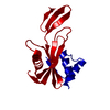 1v8iC 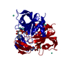 1v8mC 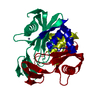 1v8nC 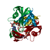 1v8rC 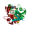 1v8sC  1v8tC 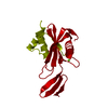 1v8uC 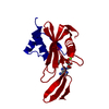 1v8vC  1v8wC 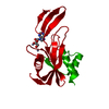 1v8yC C: citing same article ( |
|---|---|
| Similar structure data | |
| Other databases |


-Links
 PDBj
PDBj


- Assembly
| Deposited unit | 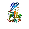
| ||||||||
|---|---|---|---|---|---|---|---|---|---|
| 1 | 
| ||||||||
| Unit cell |
|
-Components
| #1: Protein | Mass: 19287.912 Da / Num. of mol.: 1 Source method: isolated from a genetically manipulated source Source: (gene. exp.) Thermus thermophilus (bacteria) / Plasmid: pET11a / Species (production host): Escherichia coli / Production host: |
|---|---|
| #2: Chemical | ChemComp-APR /   Mass: 559.316 Da / Num. of mol.: 1 / Source method: obtained synthetically / Formula: C15H23N5O14P2 Mass: 559.316 Da / Num. of mol.: 1 / Source method: obtained synthetically / Formula: C15H23N5O14P2 |
| #3: Water | ChemComp-HOH /  Mass: 18.015 Da / Num. of mol.: 49 / Source method: isolated from a natural source / Formula: H2O Mass: 18.015 Da / Num. of mol.: 49 / Source method: isolated from a natural source / Formula: H2O |
-Experimental details
-Experiment
| Experiment | Method: X-RAY DIFFRACTION / Number of used crystals: 1 |
|---|
- Sample preparation
| Crystal | Density Matthews: 2.28 Å3/Da / Density % sol: 45.53 % |
|---|
-Data collection
| Diffraction source | Wavelength: 1.54 Å |
|---|---|
| Detector | Type: RIGAKU RAXIS IV / Detector: IMAGE PLATE |
| Radiation | Protocol: SINGLE WAVELENGTH / Monochromatic (M) / Laue (L): M / Scattering type: x-ray |
| Radiation wavelength | Wavelength: 1.54 Å / Relative weight: 1 |
| Reflection | Resolution: 2.01→20 Å / Num. obs: 11986 / % possible obs: 99.9 % / Observed criterion σ(I): 5 / Biso Wilson estimate: 8.7 Å2 / Rsym value: 0.04 |
| Reflection shell | Resolution: 2.01→2.12 Å / % possible all: 99.9 |
- Processing
| Software |
| ||||||||||||||||||||||||||||||||||||||||||||||||||||||||||||||||||||||||||||||||
|---|---|---|---|---|---|---|---|---|---|---|---|---|---|---|---|---|---|---|---|---|---|---|---|---|---|---|---|---|---|---|---|---|---|---|---|---|---|---|---|---|---|---|---|---|---|---|---|---|---|---|---|---|---|---|---|---|---|---|---|---|---|---|---|---|---|---|---|---|---|---|---|---|---|---|---|---|---|---|---|---|---|
| Refinement | Method to determine structure: MOLECULAR REPLACEMENT / Resolution: 2.1→16.14 Å / Rfactor Rfree error: 0.007 / Data cutoff high absF: 1562121.94 / Data cutoff low absF: 0 / Isotropic thermal model: RESTRAINED / Cross valid method: THROUGHOUT / σ(F): 0
| ||||||||||||||||||||||||||||||||||||||||||||||||||||||||||||||||||||||||||||||||
| Solvent computation | Solvent model: FLAT MODEL / Bsol: 56.1279 Å2 / ksol: 0.451683 e/Å3 | ||||||||||||||||||||||||||||||||||||||||||||||||||||||||||||||||||||||||||||||||
| Displacement parameters | Biso mean: 18.1 Å2
| ||||||||||||||||||||||||||||||||||||||||||||||||||||||||||||||||||||||||||||||||
| Refine analyze |
| ||||||||||||||||||||||||||||||||||||||||||||||||||||||||||||||||||||||||||||||||
| Refinement step | Cycle: LAST / Resolution: 2.1→16.14 Å
| ||||||||||||||||||||||||||||||||||||||||||||||||||||||||||||||||||||||||||||||||
| Refine LS restraints |
| ||||||||||||||||||||||||||||||||||||||||||||||||||||||||||||||||||||||||||||||||
| LS refinement shell | Resolution: 2.01→2.14 Å / Rfactor Rfree error: 0.048 / Total num. of bins used: 6
| ||||||||||||||||||||||||||||||||||||||||||||||||||||||||||||||||||||||||||||||||
| Xplor file |
|
