 Movie
Movie Controller

[English] 日本語
 Yorodumi
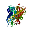
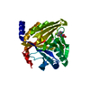
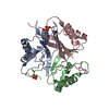
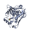
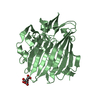
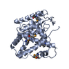
- PDB-6bnx: Crystal structure of V278E-glyoxalase I mutant from Zea mays in s... -
+ Open data

- Basic information
| Entry | Database: PDB / ID: 6bnx | ||||||
|---|---|---|---|---|---|---|---|
| Title | Crystal structure of V278E-glyoxalase I mutant from Zea mays in space group P6(3) | ||||||
 Components Components | Lactoylglutathione lyase | ||||||
 Keywords Keywords | PLANT PROTEIN / PLANT PROTEIN defense | ||||||
| Function / homology |  Function and homology information Function and homology informationlactoylglutathione lyase / lactoylglutathione lyase activity / : / metal ion binding Similarity search - Function | ||||||
| Biological species |  | ||||||
| Method |  X-RAY DIFFRACTION / SYNCHROTRON / MOLECULAR REPLACEMENT / Resolution: 1.8 Å X-RAY DIFFRACTION / SYNCHROTRON / MOLECULAR REPLACEMENT / Resolution: 1.8 Å | ||||||
 Authors Authors | Alvarez, C.E. / Agostini, R.B. / Gonzalez, J.M. / Drincovich, M.F. / Campos Bermudez, V.A. / Klinke, S. | ||||||
 Citation Citation | Journal: Febs J. / Year: 2019 Title: Deciphering the number and location of active sites in the monomeric glyoxalase I of Zea mays. Authors: Gonzalez, J.M. / Agostini, R.B. / Alvarez, C.E. / Klinke, S. / Andreo, C.S. / Campos-Bermudez, V.A. | ||||||
| History |
|
- Structure visualization
| Structure viewer | Molecule: MolmilJmol/JSmol |
|---|
- Downloads & links
-Download
| PDBx/mmCIF format | 6bnx.cif.gz | 75.8 KB | Display | PDBx/mmCIF format |
|---|---|---|---|---|
| PDB format | pdb6bnx.ent.gz | 53.5 KB | Display | PDB format |
| PDBx/mmJSON format | 6bnx.json.gz | Tree view | PDBx/mmJSON format | |
| Others |  Other downloads Other downloads |
-Validation report
| Arichive directory | https://data.pdbj.org/pub/pdb/validation_reports/bn/6bnxftp://data.pdbj.org/pub/pdb/validation_reports/bn/6bnx | HTTPS FTP |
|---|
-Related structure data
| Related structure data |  6bnnC 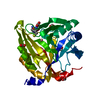 6bnzC  5d7zS S: Starting model for refinement C: citing same article ( |
|---|---|
| Similar structure data |


-Links
 PDBj
PDBj
- Assembly
| Deposited unit | 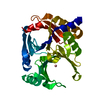
| ||||||||
|---|---|---|---|---|---|---|---|---|---|
| 1 |
| ||||||||
| Unit cell |
|
-Components
| #1: Protein | Mass: 32989.395 Da / Num. of mol.: 1 / Fragment: residues 26-315 / Mutation: V278E Source method: isolated from a genetically manipulated source Source: (gene. exp.)  |
|---|---|
| #2: Chemical | ChemComp-CO /   Mass: 58.933 Da / Num. of mol.: 1 / Source method: obtained synthetically / Formula: Co / Feature type: SUBJECT OF INVESTIGATION Mass: 58.933 Da / Num. of mol.: 1 / Source method: obtained synthetically / Formula: Co / Feature type: SUBJECT OF INVESTIGATION |
| #3: Water | ChemComp-HOH /  Mass: 18.015 Da / Num. of mol.: 154 / Source method: isolated from a natural source / Formula: H2O Mass: 18.015 Da / Num. of mol.: 154 / Source method: isolated from a natural source / Formula: H2O |
-Experimental details
-Experiment
| Experiment | Method: X-RAY DIFFRACTION / Number of used crystals: 1 |
|---|
- Sample preparation
| Crystal | Density Matthews: 2.25 Å3/Da / Density % sol: 45.35 % / Description: needle bunch |
|---|---|
| Crystal grow | Temperature: 294 K / Method: vapor diffusion, hanging drop / pH: 9.7 / Details: sodium formate 4.2 M pH 9.7, 0.5% PEG 4000 |
-Data collection
| Diffraction | Mean temperature: 100 K |
|---|---|
| Diffraction source | Source: SYNCHROTRON / Site: SOLEIL  / Beamline: PROXIMA 2 / Wavelength: 0.9801 Å / Beamline: PROXIMA 2 / Wavelength: 0.9801 Å |
| Detector | Type: DECTRIS EIGER X 9M / Detector: PIXEL / Date: Dec 11, 2016 / Details: Kirkpatrick-Baez pair of bi-morph mirrors |
| Radiation | Monochromator: channel cut cryogenically cooled monochromator crystal Protocol: SINGLE WAVELENGTH / Monochromatic (M) / Laue (L): M / Scattering type: x-ray |
| Radiation wavelength | Wavelength: 0.9801 Å / Relative weight: 1 |
| Reflection | Resolution: 1.8→41.06 Å / Num. obs: 26692 / % possible obs: 99.9 % / Redundancy: 10.27 % / CC1/2: 1 / Rrim(I) all: 0.067 / Net I/σ(I): 19.7 |
| Reflection shell | Resolution: 1.8→1.91 Å / Redundancy: 10.32 % / Mean I/σ(I) obs: 1.4 / Num. unique obs: 4293 / CC1/2: 0.666 / Rrim(I) all: 1.375 / % possible all: 99.5 |
- Processing
| Software |
| ||||||||||||||||||||||||||||||||||||||||||||||||||||||||||||||||||||||||||||||||||||||||||||||||||||||||||||||||||||||||||||||||||||||||||||||||||||||||||||||||||||||||||||||||||||||
|---|---|---|---|---|---|---|---|---|---|---|---|---|---|---|---|---|---|---|---|---|---|---|---|---|---|---|---|---|---|---|---|---|---|---|---|---|---|---|---|---|---|---|---|---|---|---|---|---|---|---|---|---|---|---|---|---|---|---|---|---|---|---|---|---|---|---|---|---|---|---|---|---|---|---|---|---|---|---|---|---|---|---|---|---|---|---|---|---|---|---|---|---|---|---|---|---|---|---|---|---|---|---|---|---|---|---|---|---|---|---|---|---|---|---|---|---|---|---|---|---|---|---|---|---|---|---|---|---|---|---|---|---|---|---|---|---|---|---|---|---|---|---|---|---|---|---|---|---|---|---|---|---|---|---|---|---|---|---|---|---|---|---|---|---|---|---|---|---|---|---|---|---|---|---|---|---|---|---|---|---|---|---|---|
| Refinement | Method to determine structure: MOLECULAR REPLACEMENT Starting model: 5D7Z Resolution: 1.8→41.05 Å / Cor.coef. Fo:Fc: 0.973 / Cor.coef. Fo:Fc free: 0.961 / SU B: 3.8 / SU ML: 0.109 / Cross valid method: THROUGHOUT / ESU R: 0.13 / ESU R Free: 0.126
| ||||||||||||||||||||||||||||||||||||||||||||||||||||||||||||||||||||||||||||||||||||||||||||||||||||||||||||||||||||||||||||||||||||||||||||||||||||||||||||||||||||||||||||||||||||||
| Solvent computation | Ion probe radii: 0.8 Å / Shrinkage radii: 0.8 Å / VDW probe radii: 1.2 Å | ||||||||||||||||||||||||||||||||||||||||||||||||||||||||||||||||||||||||||||||||||||||||||||||||||||||||||||||||||||||||||||||||||||||||||||||||||||||||||||||||||||||||||||||||||||||
| Displacement parameters | Biso mean: 38.629 Å2
| ||||||||||||||||||||||||||||||||||||||||||||||||||||||||||||||||||||||||||||||||||||||||||||||||||||||||||||||||||||||||||||||||||||||||||||||||||||||||||||||||||||||||||||||||||||||
| Refinement step | Cycle: 1 / Resolution: 1.8→41.05 Å
| ||||||||||||||||||||||||||||||||||||||||||||||||||||||||||||||||||||||||||||||||||||||||||||||||||||||||||||||||||||||||||||||||||||||||||||||||||||||||||||||||||||||||||||||||||||||
| Refine LS restraints |
|