 Movie
Movie Controller

[English] 日本語
 Yorodumi
- PDB-1v5x: Crystal structure of Phosphoribosyl anthranilate isomerase from T... -
+ Open data

- Basic information
| Entry | Database: PDB / ID: 1v5x | ||||||
|---|---|---|---|---|---|---|---|
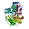
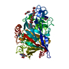
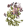
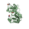
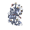
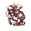
| Title | Crystal structure of Phosphoribosyl anthranilate isomerase from Thermus Thermophilus | ||||||
 Components Components | Phosphoribosylanthranilate isomerase | ||||||
 Keywords Keywords | ISOMERASE / alpha-beta barrel / TrpF / phosphoribosyl anthranilate isomerase / RIKEN Structural Genomics/Proteomics Initiative / RSGI / Structural Genomics | ||||||
| Function / homology |  Function and homology information Function and homology informationphosphoribosylanthranilate isomerase / phosphoribosylanthranilate isomerase activity / L-tryptophan biosynthetic process Similarity search - Function | ||||||
| Biological species |   Thermus thermophilus (bacteria) Thermus thermophilus (bacteria) | ||||||
| Method |  X-RAY DIFFRACTION / SYNCHROTRON / MOLECULAR REPLACEMENT / Resolution: 2 Å X-RAY DIFFRACTION / SYNCHROTRON / MOLECULAR REPLACEMENT / Resolution: 2 Å | ||||||
 Authors Authors | Taka, J. / Kunishima, N. / Yutani, K. / RIKEN Structural Genomics/Proteomics Initiative (RSGI) | ||||||
 Citation Citation | Journal: J.Biochem.(Tokyo) / Year: 2005 Title: Stabilization due to dimer formation of phosphoribosyl anthranilate isomerase from Thermus thermophilus HB8: X-ray Analysis and DSC experiments. Authors: Taka, J. / Ogasahara, K. / Jeyakanthan, J. / Kunishima, N. / Kuroishi, C. / Sugahara, M. / Yokoyama, S. / Yutani, K. | ||||||
| History |
|
- Structure visualization
| Structure viewer | Molecule: MolmilJmol/JSmol |
|---|
- Downloads & links
-Download
| PDBx/mmCIF format | 1v5x.cif.gz | 94 KB | Display | PDBx/mmCIF format |
|---|---|---|---|---|
| PDB format | pdb1v5x.ent.gz | 72.6 KB | Display | PDB format |
| PDBx/mmJSON format | 1v5x.json.gz | Tree view | PDBx/mmJSON format | |
| Others |  Other downloads Other downloads |
-Validation report
| Arichive directory | https://data.pdbj.org/pub/pdb/validation_reports/v5/1v5xftp://data.pdbj.org/pub/pdb/validation_reports/v5/1v5x | HTTPS FTP |
|---|
-Related structure data
| Related structure data |  1nsjS S: Starting model for refinement |
|---|---|
| Similar structure data | |
| Other databases |


-Links
 PDBj
PDBj
- Assembly
| Deposited unit | 
| ||||||||
|---|---|---|---|---|---|---|---|---|---|
| 1 |
| ||||||||
| Unit cell |
| ||||||||
| Details | The biological assembly is a dimer generated in the asymmetric unit. |
-Components
| #1: Protein | Mass: 21977.461 Da / Num. of mol.: 2 Source method: isolated from a genetically manipulated source Source: (gene. exp.) Thermus thermophilus (bacteria) / Plasmid: pET11a / Production host: References: UniProt: P83825, phosphoribosylanthranilate isomerase #2: Water | ChemComp-HOH / |  Mass: 18.015 Da / Num. of mol.: 301 / Source method: isolated from a natural source / Formula: H2O Mass: 18.015 Da / Num. of mol.: 301 / Source method: isolated from a natural source / Formula: H2O |
|---|
-Experimental details
-Experiment
| Experiment | Method: X-RAY DIFFRACTION / Number of used crystals: 1 |
|---|
- Sample preparation
| Crystal | Density Matthews: 2.92 Å3/Da / Density % sol: 57.52 % | ||||||||||||||||||||||||||||||||||||||||||
|---|---|---|---|---|---|---|---|---|---|---|---|---|---|---|---|---|---|---|---|---|---|---|---|---|---|---|---|---|---|---|---|---|---|---|---|---|---|---|---|---|---|---|---|
| Crystal grow | Temperature: 293 K / Method: microbatch / pH: 5.1 Details: Isopropanol, Sodium Acetate, pH 5.1, microbatch, temperature 293K | ||||||||||||||||||||||||||||||||||||||||||
| Crystal grow | *PLUS Temperature: 22 ℃ / pH: 8 / Method: batch method | ||||||||||||||||||||||||||||||||||||||||||
| Components of the solutions | *PLUS
|
-Data collection
| Diffraction | Mean temperature: 100 K |
|---|---|
| Diffraction source | Source: SYNCHROTRON / Site: SPring-8  / Beamline: BL26B1 / Wavelength: 1 Å / Beamline: BL26B1 / Wavelength: 1 Å |
| Detector | Type: RIGAKU RAXIS V / Detector: IMAGE PLATE / Date: Oct 3, 2003 / Details: mirrors |
| Radiation | Monochromator: Si 111 / Protocol: SINGLE WAVELENGTH / Monochromatic (M) / Laue (L): M / Scattering type: x-ray |
| Radiation wavelength | Wavelength: 1 Å / Relative weight: 1 |
| Reflection | Resolution: 2→35 Å / Num. all: 35156 / Num. obs: 35156 / % possible obs: 99.8 % / Observed criterion σ(F): 0 / Observed criterion σ(I): 0 / Redundancy: 8.7 % / Biso Wilson estimate: 29.43 Å2 / Rmerge(I) obs: 0.08 / Rsym value: 0.076 / Net I/σ(I): 9.92 |
| Reflection shell | Resolution: 2→2.07 Å / Redundancy: 5 % / Rmerge(I) obs: 0.763 / Mean I/σ(I) obs: 3 / Num. unique all: 3404 / Rsym value: 0.698 / % possible all: 99.4 |
| Reflection | *PLUS Num. measured all: 307402 / Rmerge(I) obs: 0.08 |
| Reflection shell | *PLUS % possible obs: 99.4 % |
- Processing
| Software |
| |||||||||||||||||||||||||
|---|---|---|---|---|---|---|---|---|---|---|---|---|---|---|---|---|---|---|---|---|---|---|---|---|---|---|
| Refinement | Method to determine structure: MOLECULAR REPLACEMENT Starting model: PDB entry 1NSJ Resolution: 2→35 Å / Isotropic thermal model: Anisotropic / Cross valid method: THROUGHOUT / σ(F): 0 / Stereochemistry target values: Engh & Huber
| |||||||||||||||||||||||||
| Displacement parameters | Biso mean: 44.9 Å2
| |||||||||||||||||||||||||
| Refine analyze |
| |||||||||||||||||||||||||
| Refinement step | Cycle: LAST / Resolution: 2→35 Å
| |||||||||||||||||||||||||
| Refine LS restraints |
| |||||||||||||||||||||||||
| LS refinement shell | Resolution: 2→2.09 Å / Rfactor Rfree error: 0.026
| |||||||||||||||||||||||||
| Refinement | *PLUS % reflection Rfree: 5 % | |||||||||||||||||||||||||
| Solvent computation | *PLUS | |||||||||||||||||||||||||
| Displacement parameters | *PLUS |
