 Movie
Movie Controller

[English] 日本語
 Yorodumi
- PDB-1v4n: Structure of 5'-deoxy-5'-methylthioadenosine phosphorylase homolo... -
+ Open data

- Basic information
| Entry | Database: PDB / ID: 1v4n | ||||||
|---|---|---|---|---|---|---|---|
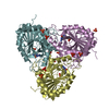
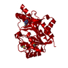
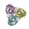
| Title | Structure of 5'-deoxy-5'-methylthioadenosine phosphorylase homologue from Sulfolobus tokodaii | ||||||
 Components Components | 271aa long hypothetical 5'-methylthioadenosine phosphorylase | ||||||
 Keywords Keywords | TRANSFERASE / STRUCTURAL GENOMICS | ||||||
| Function / homology |  Function and homology information Function and homology informationS-methyl-5'-thioadenosine phosphorylase / S-methyl-5-thioadenosine phosphorylase activity / : / purine ribonucleoside salvage / cytosol Similarity search - Function | ||||||
| Biological species |   Sulfolobus tokodaii (archaea) Sulfolobus tokodaii (archaea) | ||||||
| Method |  X-RAY DIFFRACTION / SYNCHROTRON / MOLECULAR REPLACEMENT / Resolution: 2.45 Å X-RAY DIFFRACTION / SYNCHROTRON / MOLECULAR REPLACEMENT / Resolution: 2.45 Å | ||||||
 Authors Authors | Kitago, Y. / Yasutake, Y. / Sakai, N. / Tsujimura, M. / Yao, M. / Watanabe, N. / Kawarabayasi, Y. / Tanaka, I. | ||||||
 Citation Citation | Journal: To be Published Title: Crystal structure of Sulfolobus tokodaii MTAP Authors: Kitago, Y. / Yasutake, Y. / Sakai, N. / Tsujimura, M. / Yao, M. / Watanabe, N. / Kawarabayasi, Y. / Tanaka, I. | ||||||
| History |
|
- Structure visualization
| Structure viewer | Molecule: MolmilJmol/JSmol |
|---|
- Downloads & links
-Download
| PDBx/mmCIF format | 1v4n.cif.gz | 166.5 KB | Display | PDBx/mmCIF format |
|---|---|---|---|---|
| PDB format | pdb1v4n.ent.gz | 134.7 KB | Display | PDB format |
| PDBx/mmJSON format | 1v4n.json.gz | Tree view | PDBx/mmJSON format | |
| Others |  Other downloads Other downloads |
-Validation report
| Arichive directory | https://data.pdbj.org/pub/pdb/validation_reports/v4/1v4nftp://data.pdbj.org/pub/pdb/validation_reports/v4/1v4n | HTTPS FTP |
|---|
-Related structure data
| Related structure data |  1cg6S S: Starting model for refinement |
|---|---|
| Similar structure data |


-Links
 PDBj
PDBj- Assembly
| Deposited unit | 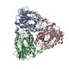
| ||||||||
|---|---|---|---|---|---|---|---|---|---|
| 1 |
| ||||||||
| Unit cell |
| ||||||||
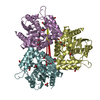
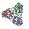
| Details | The biological assembly is homotrimer in the asymmetric unit. |
-Components
| #1: Protein | Mass: 31582.643 Da / Num. of mol.: 3 Source method: isolated from a genetically manipulated source Source: (gene. exp.) Sulfolobus tokodaii (archaea) / Gene: ST0485 / Plasmid: pET-23a / Production host:  References: GenBank: 15920700, UniProt: F9VN03*PLUS, S-methyl-5'-thioadenosine phosphorylase #2: Water | ChemComp-HOH / |  Mass: 18.015 Da / Num. of mol.: 275 / Source method: isolated from a natural source / Formula: H2O Mass: 18.015 Da / Num. of mol.: 275 / Source method: isolated from a natural source / Formula: H2OHas protein modification | Y | |
|---|
-Experimental details
-Experiment
| Experiment | Method: X-RAY DIFFRACTION / Number of used crystals: 1 |
|---|
- Sample preparation
| Crystal | Density Matthews: 2.64 Å3/Da / Density % sol: 53.08 % |
|---|---|
| Crystal grow | Temperature: 293 K / Method: vapor diffusion, sitting drop / pH: 7 Details: Tris buffer, PEG1000, pH 7.0, VAPOR DIFFUSION, SITTING DROP, temperature 293K |
-Data collection
| Diffraction | Mean temperature: 100 K |
|---|---|
| Diffraction source | Source: SYNCHROTRON / Site: Photon Factory  / Beamline: BL-18B / Wavelength: 1 Å / Beamline: BL-18B / Wavelength: 1 Å |
| Detector | Type: ADSC QUANTUM 4 / Detector: CCD / Date: May 27, 2003 / Details: a mirror and monochrometer |
| Radiation | Monochromator: Si 111 / Protocol: SINGLE WAVELENGTH / Monochromatic (M) / Laue (L): M / Scattering type: x-ray |
| Radiation wavelength | Wavelength: 1 Å / Relative weight: 1 |
| Reflection | Resolution: 2.45→50 Å / Num. all: 36084 / Num. obs: 36084 / % possible obs: 100 % / Observed criterion σ(I): -3 / Redundancy: 6.93 % / Biso Wilson estimate: 33.59 Å2 / Rsym value: 0.101 / Net I/σ(I): 13.7 |
| Reflection shell | Resolution: 2.45→2.54 Å / Redundancy: 6.81 % / Rmerge(I) obs: 0.229 / Num. unique all: 3576 / Rsym value: 0.229 / % possible all: 100 |
- Processing
| Software |
| |||||||||||||||||||||||||||
|---|---|---|---|---|---|---|---|---|---|---|---|---|---|---|---|---|---|---|---|---|---|---|---|---|---|---|---|---|
| Refinement | Method to determine structure: MOLECULAR REPLACEMENT Starting model: PDB code 1cg6 Resolution: 2.45→50 Å / Isotropic thermal model: isotropic / Cross valid method: THROUGHOUT / σ(F): 0 / Stereochemistry target values: Engh & Huber
| |||||||||||||||||||||||||||
| Displacement parameters | Biso mean: 34.228 Å2
| |||||||||||||||||||||||||||
| Refine analyze |
| |||||||||||||||||||||||||||
| Refinement step | Cycle: LAST / Resolution: 2.45→50 Å
| |||||||||||||||||||||||||||
| Refine LS restraints |
| |||||||||||||||||||||||||||
| LS refinement shell | Resolution: 2.45→2.54 Å / Total num. of bins used: 10
|
