[English] 日本語
Yorodumi
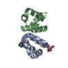
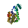
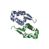
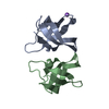
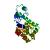
- PDB-1uv7: periplasmic domain of EpsM from Vibrio cholerae -

+
Open data


ID or keywords:

Loading...

-
Basic information

Entry
Database: PDB / ID: 1uv7
Titleperiplasmic domain of EpsM from Vibrio cholerae
ComponentsGENERAL SECRETION PATHWAY PROTEIN M
KeywordsTRANSPORT / GENERAL SECRETION PATHWAY / VIBRIO CHOLERAE
Function / homology
Function and homology information


protein secretion by the type II secretion system / type II protein secretion system complex / : / membrane => GO:0016020 / plasma membrane
Similarity search - Function
Type II secretion system protein GspM / Type II secretion system protein GspM, periplasmic domain superfamily / Type II secretion system (T2SS), protein M / General secretion pathway protein M, EpsM / Gyrase A; domain 2 / 2-Layer Sandwich / Alpha Beta
Similarity search - Domain/homology
Type II secretion system protein M
Similarity search - Component
Biological speciesVIBRIO CHOLERAE (bacteria)
MethodX-RAY DIFFRACTION / SYNCHROTRON / MAD / Resolution: 1.7 Å
AuthorsAbendroth, J. / Hol, W.G.J.
CitationJournal: J.Mol.Biol. / Year: 2004
Title: The Crystal Structure of the Periplasmic Domain of the Type II Secretion System Protein Epsm from Vibrio Cholerae: The Simplest Version of the Ferredoxin Fold
Authors: Abendroth, J. / Rice, A. / Mcluskey, K. / Bagdasarian, M. / Hol, W.G.J.
History
DepositionJan 15, 2004Deposition site: PDBE / Processing site: PDBE
Revision 1.0Apr 23, 2004Provider: repository / Type: Initial release
Revision 1.1Jul 13, 2011Group: Advisory / Version format compliance
Revision 1.2May 15, 2019Group: Data collection / Derived calculations ...Data collection / Derived calculations / Experimental preparation / Other
Category: exptl_crystal_grow / pdbx_database_proc ...exptl_crystal_grow / pdbx_database_proc / pdbx_database_status / struct_conn
Item: _exptl_crystal_grow.temp / _pdbx_database_status.recvd_author_approval / _struct_conn.pdbx_leaving_atom_flag

-
Structure visualization

Structure viewerMolecule:
MolmilJmol/JSmol

Downloads & links

-
Assembly

Deposited unit
A: GENERAL SECRETION PATHWAY PROTEIN M
B: GENERAL SECRETION PATHWAY PROTEIN M


Theoretical massNumber of molelcules
Total (without water)25,2802
Polymers25,2802
Non-polymers00
Water1,26170
1


  • Idetical with deposited unit
  • defined by author&software
TypeNameSymmetry operationNumber
identity operation1_555x,y,z1
MethodPQS
Unit cell
Length a, b, c (Å)52.879, 52.879, 112.484
Angle α, β, γ (deg.)90.00, 90.00, 120.00
Int Tables number154
Space group name H-MP3221
Noncrystallographic symmetry (NCS)NCS oper: (Code: given
Matrix: (-0.60938, -0.79177, 0.04179), (-0.792, 0.6054, -0.07888), (0.03716, -0.08117, -0.99601)
Vector: 78.71606, 39.90469, 18.97545)

-
Components

#1: Protein GENERAL SECRETION PATHWAY PROTEIN M / CHOLERA TOXIN SECRETION PROTEIN EPSM


Mass: 12639.798 Da / Num. of mol.: 2 / Fragment: PERIPLASMIC DOMAIN, RESIDUES 65-165
Source method: isolated from a genetically manipulated source
Source: (gene. exp.) VIBRIO CHOLERAE (bacteria) / Plasmid: PET21D(+) / Production host: ESCHERICHIA COLI (E. coli) / Strain (production host): BL21(DE3) / References: UniProt: P41851
#2: Water ChemComp-HOH / water


Mass: 18.015 Da / Num. of mol.: 70 / Source method: isolated from a natural source / Formula: H2O
Compound detailsREQUIRED FOR SECRETION OF CHOLERA TOXIN THROUGH THE OUTER MEMBRANE.
Sequence detailsRESIDUES 166 TO 173 IN THE SEQRES RECORDS GIVEN BELOW ARE FROM THE HIS-TAG USED FOR THE EXPRESSION ...RESIDUES 166 TO 173 IN THE SEQRES RECORDS GIVEN BELOW ARE FROM THE HIS-TAG USED FOR THE EXPRESSION OF THE PROTEIN. RESIDUE 64 IS THE INITIAL EXPRESSION METHIONINE RESIDUE.

-
Experimental details

-
Experiment

ExperimentMethod: X-RAY DIFFRACTION / Number of used crystals: 1

-
Sample preparation

CrystalDensity Matthews: 1.8 Å3/Da / Density % sol: 32.4 %
Crystal growTemperature: 277 K / pH: 8
Details: 6-12MG/ML PROTEIN IN 20MM TRIS PH8, 150MM NACL, 1MM TCEP; RESERVOIR: 2.4-3.0M SODIUM MALONATE, 100MM TRIS PH~8; CRYSTALLISATION: 1.5MKL PROTEIN + 1.5MKL RESERVIOR, 4C, pH 8.00

-
Data collection

DiffractionMean temperature: 100 K
Diffraction sourceSource: SYNCHROTRON / Site: ALS / Beamline: 8.2.2 / Wavelength: 0.9791,0.9795,0.9686
DetectorType: ADSC CCD / Detector: CCD / Date: Dec 12, 2002
RadiationProtocol: MAD / Monochromatic (M) / Laue (L): M / Scattering type: x-ray
Radiation wavelength
IDWavelength (Å)Relative weight
10.97911
20.97951
30.96861
ReflectionResolution: 1.7→15.29 Å / Num. obs: 20676 / % possible obs: 99.7 % / Redundancy: 6.61 % / Rmerge(I) obs: 0.047 / Net I/σ(I): 18
Reflection shellResolution: 1.7→1.79 Å / Redundancy: 5.5 % / Rmerge(I) obs: 0.627 / Mean I/σ(I) obs: 2.6 / % possible all: 93.4

-
Processing

Software
NameVersionClassification
REFMAC5.1.24refinement
MOSFLMdata reduction
SCALAdata scaling
SOLVEphasing
RefinementMethod to determine structure: MAD / Resolution: 1.7→20 Å / Cor.coef. Fo:Fc: 0.96 / Cor.coef. Fo:Fc free: 0.945 / SU B: 2.489 / SU ML: 0.081 / TLS residual ADP flag: LIKELY RESIDUAL / Cross valid method: THROUGHOUT / ESU R: 0.103 / ESU R Free: 0.108 / Stereochemistry target values: MAXIMUM LIKELIHOOD
Details: HYDROGENS HAVE BEEN ADDED IN THE RIDING POSITIONS. N-TERM 65-85 DISORDERED, LOOP 149-152 DISORDERED, HIS6-TAG DISORDERED
RfactorNum. reflection% reflectionSelection details
Rfree0.242 1027 5 %RANDOM
Rwork0.196 ---
obs-19604 99.6 %-
Solvent computationIon probe radii: 0.8 Å / Shrinkage radii: 0.8 Å / VDW probe radii: 1.4 Å / Solvent model: BABINET MODEL PLUS MASK
Displacement parametersBiso mean: 21.95 Å2
Baniso -1Baniso -2Baniso -3
1--0.42 Å2-0.21 Å20 Å2
2---0.42 Å20 Å2
3---0.63 Å2
Refinement stepCycle: LAST / Resolution: 1.7→20 Å
ProteinNucleic acidLigandSolventTotal
Num. atoms1228 0 0 70 1298
Refine LS restraints
Refine-IDTypeDev idealDev ideal targetNumber
X-RAY DIFFRACTIONr_bond_refined_d0.0140.0221262
X-RAY DIFFRACTIONr_bond_other_d0.0020.021216
X-RAY DIFFRACTIONr_angle_refined_deg2.3021.9481704
X-RAY DIFFRACTIONr_angle_other_deg1.06832820
X-RAY DIFFRACTIONr_dihedral_angle_1_deg5.4695146
X-RAY DIFFRACTIONr_dihedral_angle_2_deg
X-RAY DIFFRACTIONr_dihedral_angle_3_deg
X-RAY DIFFRACTIONr_dihedral_angle_4_deg
X-RAY DIFFRACTIONr_chiral_restr0.1580.2196
X-RAY DIFFRACTIONr_gen_planes_refined0.0120.021344
X-RAY DIFFRACTIONr_gen_planes_other0.0010.02242
X-RAY DIFFRACTIONr_nbd_refined0.2340.2236
X-RAY DIFFRACTIONr_nbd_other0.2530.21415
X-RAY DIFFRACTIONr_nbtor_refined
X-RAY DIFFRACTIONr_nbtor_other0.090.2885
X-RAY DIFFRACTIONr_xyhbond_nbd_refined0.2140.246
X-RAY DIFFRACTIONr_xyhbond_nbd_other
X-RAY DIFFRACTIONr_metal_ion_refined
X-RAY DIFFRACTIONr_metal_ion_other
X-RAY DIFFRACTIONr_symmetry_vdw_refined0.1590.221
X-RAY DIFFRACTIONr_symmetry_vdw_other0.3050.243
X-RAY DIFFRACTIONr_symmetry_hbond_refined0.1780.212
X-RAY DIFFRACTIONr_symmetry_hbond_other
X-RAY DIFFRACTIONr_symmetry_metal_ion_refined
X-RAY DIFFRACTIONr_symmetry_metal_ion_other
X-RAY DIFFRACTIONr_mcbond_it1.521.5750
X-RAY DIFFRACTIONr_mcbond_other
X-RAY DIFFRACTIONr_mcangle_it2.721232
X-RAY DIFFRACTIONr_mcangle_other
X-RAY DIFFRACTIONr_scbond_it4.7063512
X-RAY DIFFRACTIONr_scbond_other
X-RAY DIFFRACTIONr_scangle_it6.9264.5472
X-RAY DIFFRACTIONr_scangle_other
X-RAY DIFFRACTIONr_long_range_B_refined
X-RAY DIFFRACTIONr_long_range_B_other
X-RAY DIFFRACTIONr_rigid_bond_restr
X-RAY DIFFRACTIONr_sphericity_free
X-RAY DIFFRACTIONr_sphericity_bonded
LS refinement shellResolution: 1.7→1.74 Å / Total num. of bins used: 20 /
RfactorNum. reflection
Rfree0.274 62
Rwork0.262 1410
Refinement TLS params.

Method: refined / Refine-ID: X-RAY DIFFRACTION

IDL112)L122)L132)L222)L232)L332)S11 (Å °)S12 (Å °)S13 (Å °)S21 (Å °)S22 (Å °)S23 (Å °)S31 (Å °)S32 (Å °)S33 (Å °)T112)T122)T132)T222)T232)T332)Origin x (Å)Origin y (Å)Origin z (Å)
15.23730.885-2.69845.43392.98327.0751-0.0268-0.10020.32890.0729-0.05850.62170.0438-0.43460.08530.03970.00490.03760.12420.02290.124828.0124.960316.6642
25.41773.37340.36366.4544-2.30035.6229-0.29420.33920.0964-0.34220.2726-0.0987-0.30540.33890.02160.1114-0.07790.03830.0722-0.01360.022942.356731.46741.27
Refinement TLS group
IDRefine-IDRefine TLS-IDAuth asym-IDAuth seq-ID
1X-RAY DIFFRACTION1A86 - 165
2X-RAY DIFFRACTION2B86 - 163

+
About Yorodumi

-
News

-
Feb 9, 2022. New format data for meta-information of EMDB entries

New format data for meta-information of EMDB entries

  • Version 3 of the EMDB header file is now the official format.
  • The previous official version 1.9 will be removed from the archive.

Related info.:EMDB header

External links:wwPDB to switch to version 3 of the EMDB data model

-
Aug 12, 2020. Covid-19 info

Covid-19 info

URL: https://pdbjlvh1.pdbj.org/emnavi/covid19.php

New page: Covid-19 featured information page in EM Navigator.

Related info.:Covid-19 info / Mar 5, 2020. Novel coronavirus structure data

+
Mar 5, 2020. Novel coronavirus structure data

Novel coronavirus structure data

Related info.:Yorodumi Speices / Aug 12, 2020. Covid-19 info

External links:COVID-19 featured content - PDBj / Molecule of the Month (242):Coronavirus Proteases

+
Jan 31, 2019. EMDB accession codes are about to change! (news from PDBe EMDB page)

EMDB accession codes are about to change! (news from PDBe EMDB page)

  • The allocation of 4 digits for EMDB accession codes will soon come to an end. Whilst these codes will remain in use, new EMDB accession codes will include an additional digit and will expand incrementally as the available range of codes is exhausted. The current 4-digit format prefixed with “EMD-” (i.e. EMD-XXXX) will advance to a 5-digit format (i.e. EMD-XXXXX), and so on. It is currently estimated that the 4-digit codes will be depleted around Spring 2019, at which point the 5-digit format will come into force.
  • The EM Navigator/Yorodumi systems omit the EMD- prefix.

Related info.:Q: What is EMD? / ID/Accession-code notation in Yorodumi/EM Navigator

External links:EMDB Accession Codes are Changing Soon! / Contact to PDBj

+
Jul 12, 2017. Major update of PDB

Major update of PDB

  • wwPDB released updated PDB data conforming to the new PDBx/mmCIF dictionary.
  • This is a major update changing the version number from 4 to 5, and with Remediation, in which all the entries are updated.
  • In this update, many items about electron microscopy experimental information are reorganized (e.g. em_software).
  • Now, EM Navigator and Yorodumi are based on the updated data.

External links:wwPDB Remediation / Enriched Model Files Conforming to OneDep Data Standards Now Available in the PDB FTP Archive

-
Yorodumi

Thousand views of thousand structures

  • Yorodumi is a browser for structure data from EMDB, PDB, SASBDB, etc.
  • This page is also the successor to EM Navigator detail page, and also detail information page/front-end page for Omokage search.
  • The word "yorodu" (or yorozu) is an old Japanese word meaning "ten thousand". "mi" (miru) is to see.

Related info.:EMDB / PDB / SASBDB / Comparison of 3 databanks / Yorodumi Search / Aug 31, 2016. New EM Navigator & Yorodumi / Yorodumi Papers / Jmol/JSmol / Function and homology information / Changes in new EM Navigator and Yorodumi

Read more