 Movie
Movie Controller

[English] 日本語
 Yorodumi
- PDB-1utg: REFINEMENT OF THE C2221 CRYSTAL FORM OF OXIDIZED UTEROGLOBIN AT 1... -
+ Open data

- Basic information
| Entry | Database: PDB / ID: 1utg | ||||||
|---|---|---|---|---|---|---|---|
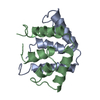
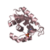
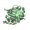
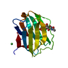
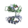
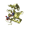
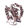
| Title | REFINEMENT OF THE C2221 CRYSTAL FORM OF OXIDIZED UTEROGLOBIN AT 1.34 ANGSTROMS RESOLUTION | ||||||
 Components Components | UTEROGLOBIN | ||||||
 Keywords Keywords | STEROID BINDING | ||||||
| Function / homology |  Function and homology information Function and homology informationphospholipase A2 inhibitor activity / steroid binding / signal transduction / : / cytoplasm Similarity search - Function | ||||||
| Biological species |  | ||||||
| Method |  X-RAY DIFFRACTION / Resolution: 1.34 Å X-RAY DIFFRACTION / Resolution: 1.34 Å | ||||||
 Authors Authors | Morize, I. / Surcouf, E. / Vaney, M.C. / Buehner, M. / Mornon, J.P. | ||||||
 Citation Citation | Journal: J.Mol.Biol. / Year: 1987 Title: Refinement of the C222(1) crystal form of oxidized uteroglobin at 1.34 A resolution. Authors: Morize, I. / Surcouf, E. / Vaney, M.C. / Epelboin, Y. / Buehner, M. / Fridlansky, F. / Milgrom, E. / Mornon, J.P. #1: Journal: FEBS Lett. / Year: 1988Title: Evidence for the Identity of Anti-Proteinase Pulmonary Protein Ccsp and Uteroglobin Authors: Lopez Deharo, M.S. / Alvarez, L. / Nieto, A. #2: Journal: Biochem.J. / Year: 1987Title: Purification, Characterization and Proteinase-Inhibitory Activity of a Clara-Cell Secretory Protein from the Pulmonary Extracellular Lining of Rabbits Authors: Gupta, R.P. / Patton, S.E. / Jetten, A.M. / Hook, G.E.R. #3: Journal: Endocr.Rev. / Year: 1987Title: Uteroglobin. Structure, Molecular Biology, and New Perspectives on its Function as a Phospholipasea2 Inhibitor Authors: Miele, L. / Cordella-Miele, E. / Mukherjee, A.B. #4: Journal: J.Mol.Biol. / Year: 1982Title: Use of Molecular Replacement in the Structure Determination of the P21212 and the P21 (Pseudo P21212) Crystal Forms of Oxidized Uteroglobin Authors: Buehner, M. / Lifchitz, A. / Bally, R. / Mornon, J.P. #5: Journal: J.Mol.Biol. / Year: 1980Title: X-Ray Crystallographic Analysis of a Progesterone-Binding Protein. The C 2 2 21 Crystal Form of Oxidized Uteroglobin at 2.2 Angstroms Resolution Authors: Mornon, J.P. / Fridlansky, F. / Bally, R. / Milgrom, E. #6: Journal: J.Mol.Biol. / Year: 1979Title: Characterization of Two New Crystal Forms of Uteroglobin Authors: Mornon, J.P. / Bally, R. / Fridlansky, F. / Milgrom, E. #7: Journal: J.Mol.Biol. / Year: 1978Title: X-Ray Analysis of a Progesterone-Binding Protein (Uteroglobin). Preliminary Results Authors: Mornon, J.P. / Surcouf, E. / Bally, R. / Fridlansky, F. / Milgrom, E. | ||||||
| History |
|
- Structure visualization
| Structure viewer | Molecule: MolmilJmol/JSmol |
|---|
- Downloads & links
-Download
| PDBx/mmCIF format | 1utg.cif.gz | 27.5 KB | Display | PDBx/mmCIF format |
|---|---|---|---|---|
| PDB format | pdb1utg.ent.gz | 17.9 KB | Display | PDB format |
| PDBx/mmJSON format | 1utg.json.gz | Tree view | PDBx/mmJSON format | |
| Others |  Other downloads Other downloads |
-Validation report
| Arichive directory | https://data.pdbj.org/pub/pdb/validation_reports/ut/1utgftp://data.pdbj.org/pub/pdb/validation_reports/ut/1utg | HTTPS FTP |
|---|
-Related structure data
| Similar structure data |
|---|

-Links
 PDBj
PDBj- Assembly
| Deposited unit | 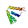
| ||||||||
|---|---|---|---|---|---|---|---|---|---|
| 1 | 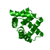
| ||||||||
| Unit cell |
| ||||||||
| Atom site foot note | 1: SEE REMARK 5. | ||||||||
| Components on special symmetry positions |
|
-Components
| #1: Protein | Mass: 7910.272 Da / Num. of mol.: 1 Source method: isolated from a genetically manipulated source Source: (gene. exp.) |
|---|---|
| #2: Water | ChemComp-HOH /  Mass: 18.015 Da / Num. of mol.: 83 / Source method: isolated from a natural source / Formula: H2O Mass: 18.015 Da / Num. of mol.: 83 / Source method: isolated from a natural source / Formula: H2O |
| Compound details | THE SECONDARY STRUCTURE CONSISTS OF FOUR ALPHA-HELICES AND ONE BETA-TURN PER MONOMER. A CAVITY ...THE SECONDARY STRUCTURE CONSISTS OF FOUR ALPHA-HELICES AND ONE BETA-TURN PER MONOMER. A CAVITY EXISTS IN THE DIMER, BETWEEN THE TWO MONOMERS. ITS LENGTH IS 15.6 ANGSTROMS AND ITS WIDTH IS 9. ANGSTROMS. 14 WATER MOLECULES POSITIONS COULD BE RECOGNISED |
| Has protein modification | Y |
-Experimental details
-Experiment
| Experiment | Method: X-RAY DIFFRACTION |
|---|
- Sample preparation
| Crystal | Density Matthews: 1.98 Å3/Da / Density % sol: 37.97 % | ||||||||||||||||||||||||||||||
|---|---|---|---|---|---|---|---|---|---|---|---|---|---|---|---|---|---|---|---|---|---|---|---|---|---|---|---|---|---|---|---|
| Crystal grow | *PLUS Method: vapor diffusion / Details: took from Mornon et al., from original paper | ||||||||||||||||||||||||||||||
| Components of the solutions | *PLUS
|
-Data collection
| Reflection | *PLUS Highest resolution: 1.34 Å / Lowest resolution: 22 Å / Num. obs: 7509 / Rmerge(I) obs: 0.089 |
|---|
- Processing
| Software | Name: PROLSQ / Classification: refinement | ||||||||||||||||||||||||||||||||||||||||||||||||||||||||||||||||||||||||||||||||||||
|---|---|---|---|---|---|---|---|---|---|---|---|---|---|---|---|---|---|---|---|---|---|---|---|---|---|---|---|---|---|---|---|---|---|---|---|---|---|---|---|---|---|---|---|---|---|---|---|---|---|---|---|---|---|---|---|---|---|---|---|---|---|---|---|---|---|---|---|---|---|---|---|---|---|---|---|---|---|---|---|---|---|---|---|---|---|
| Refinement | Rfactor obs: 0.23 / Highest resolution: 1.34 Å | ||||||||||||||||||||||||||||||||||||||||||||||||||||||||||||||||||||||||||||||||||||
| Refinement step | Cycle: LAST / Highest resolution: 1.34 Å
| ||||||||||||||||||||||||||||||||||||||||||||||||||||||||||||||||||||||||||||||||||||
| Refine LS restraints |
| ||||||||||||||||||||||||||||||||||||||||||||||||||||||||||||||||||||||||||||||||||||
| Software | *PLUS Name: PROLSQ / Classification: refinement | ||||||||||||||||||||||||||||||||||||||||||||||||||||||||||||||||||||||||||||||||||||
| Refinement | *PLUS Lowest resolution: 5 Å / Num. reflection obs: 14227 / Rfactor obs: 0.23 | ||||||||||||||||||||||||||||||||||||||||||||||||||||||||||||||||||||||||||||||||||||
| Solvent computation | *PLUS | ||||||||||||||||||||||||||||||||||||||||||||||||||||||||||||||||||||||||||||||||||||
| Displacement parameters | *PLUS |
