 Movie
Movie Controller

[English] 日本語
 Yorodumi
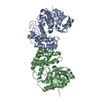
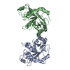
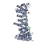
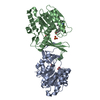
- PDB-1us2: Xylanase10C (mutant E385A) from Cellvibrio japonicus in complex w... -
+ Open data

- Basic information
| Entry | Database: PDB / ID: 1us2 | |||||||||
|---|---|---|---|---|---|---|---|---|---|---|
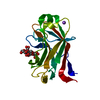
| Title | Xylanase10C (mutant E385A) from Cellvibrio japonicus in complex with xylopentaose | |||||||||
 Components Components | ENDO-BETA-1,4-XYLANASE | |||||||||
 Keywords Keywords | HYDROLASE / CARBOHYDRATE BINDING MODULE / XYLAN DEGRADATION | |||||||||
| Function / homology |  Function and homology information Function and homology informationendo-1,4-beta-xylanase / endo-1,4-beta-xylanase activity / xylan catabolic process / cell outer membrane Similarity search - Function | |||||||||
| Biological species |  CELLVIBRIO JAPONICUS (bacteria) CELLVIBRIO JAPONICUS (bacteria) | |||||||||
| Method |  X-RAY DIFFRACTION / SYNCHROTRON / MOLECULAR REPLACEMENT / Resolution: 1.85 Å X-RAY DIFFRACTION / SYNCHROTRON / MOLECULAR REPLACEMENT / Resolution: 1.85 Å | |||||||||
 Authors Authors | Pell, G. / Szabo, L. / Charnock, S.J. / Xie, H. / Gloster, T.M. / Davies, G.J. / Gilbert, H.J. | |||||||||
 Citation Citation | Journal: J.Biol.Chem. / Year: 2004 Title: Structural and Biochemical Analysis of Cellvibrio Japonicus Xylanase 10C: How Variation in Substrate-Binding Cleft Influences the Catalytic Profile of Family Gh-10 Xylanases Authors: Pell, G. / Szabo, L. / Charnock, S.J. / Xie, H. / Gloster, T.M. / Davies, G.J. / Gilbert, H.J. | |||||||||
| History |
| |||||||||
| Remark 700 | SHEET DETERMINATION METHOD: DSSP THE SHEETS PRESENTED AS "AD" IN EACH CHAIN ON SHEET RECORDS BELOW ... SHEET DETERMINATION METHOD: DSSP THE SHEETS PRESENTED AS "AD" IN EACH CHAIN ON SHEET RECORDS BELOW IS ACTUALLY AN 9-STRANDED BARREL THIS IS REPRESENTED BY A 10-STRANDED SHEET IN WHICH THE FIRST AND LAST STRANDS ARE IDENTICAL. |
- Structure visualization
| Structure viewer | Molecule: MolmilJmol/JSmol |
|---|
- Downloads & links
-Download
| PDBx/mmCIF format | 1us2.cif.gz | 136.2 KB | Display | PDBx/mmCIF format |
|---|---|---|---|---|
| PDB format | pdb1us2.ent.gz | 103.8 KB | Display | PDB format |
| PDBx/mmJSON format | 1us2.json.gz | Tree view | PDBx/mmJSON format | |
| Others |  Other downloads Other downloads |
-Validation report
| Arichive directory | https://data.pdbj.org/pub/pdb/validation_reports/us/1us2ftp://data.pdbj.org/pub/pdb/validation_reports/us/1us2 | HTTPS FTP |
|---|
-Related structure data
| Related structure data | 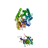 1us3C  1clxS C: citing same article ( S: Starting model for refinement |
|---|---|
| Similar structure data |


-Links
 PDBj
PDBj


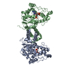
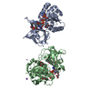
- Assembly
| Deposited unit | 
| ||||||||
|---|---|---|---|---|---|---|---|---|---|
| 1 |
| ||||||||
| Unit cell |
|
-Components
| #1: Protein | Mass: 58152.391 Da / Num. of mol.: 1 Fragment: CARBOHYDRATE BINDING MODULE AND CATALYTIC MODULE, RESIDUES (86-606) Mutation: YES Source method: isolated from a genetically manipulated source Source: (gene. exp.) CELLVIBRIO JAPONICUS (bacteria) / Production host: | ||||||||
|---|---|---|---|---|---|---|---|---|---|
| #2: Polysaccharide | Source method: isolated from a genetically manipulated source #3: Water | ChemComp-HOH / |  Mass: 18.015 Da / Num. of mol.: 833 / Source method: isolated from a natural source / Formula: H2O Mass: 18.015 Da / Num. of mol.: 833 / Source method: isolated from a natural source / Formula: H2OCompound details | ENGINEERED | Has protein modification | Y | Sequence details | NUMBERING IS CONSISTENT WITH THE SWISSPROT ENTRY FOR THE GENE. 3 MISTAKES WERE DISCOVERED IN ...NUMBERING IS CONSISTENT | |
-Experimental details
-Experiment
| Experiment | Method: X-RAY DIFFRACTION / Number of used crystals: 1 |
|---|
- Sample preparation
| Crystal | Density Matthews: 2.4 Å3/Da / Density % sol: 48.9 % | |||||||||||||||||||||||||
|---|---|---|---|---|---|---|---|---|---|---|---|---|---|---|---|---|---|---|---|---|---|---|---|---|---|---|
| Crystal grow | pH: 7 Details: 30 MG/ML PROTEIN 0.2 M SODIUM IODIDE, 20% PEG 3350, 20 MM (1 UL) XYLOPENTAOSE, pH 7.00 | |||||||||||||||||||||||||
| Crystal grow | *PLUS Temperature: 18 ℃ / Method: vapor diffusion, hanging drop | |||||||||||||||||||||||||
| Components of the solutions | *PLUS
|
-Data collection
| Diffraction | Mean temperature: 100 K |
|---|---|
| Diffraction source | Source: SYNCHROTRON / Site: SRS  / Beamline: PX9.6 / Wavelength: 0.87 / Beamline: PX9.6 / Wavelength: 0.87 |
| Detector | Type: ADSC QUANTUM 4 / Detector: CCD / Date: May 15, 2001 / Details: VERTICALLY FOCUSSING RH COATED SI MIRROR |
| Radiation | Monochromator: TRIANGULAR SINGLE CRYSTAL SI MONOCHROMATOR / Protocol: SINGLE WAVELENGTH / Monochromatic (M) / Laue (L): M / Scattering type: x-ray |
| Radiation wavelength | Wavelength: 0.87 Å / Relative weight: 1 |
| Reflection | Resolution: 1.85→20 Å / Num. obs: 53227 / % possible obs: 98 % / Redundancy: 4.4 % / Rmerge(I) obs: 0.08 / Net I/σ(I): 18.5 |
| Reflection shell | Resolution: 1.85→1.92 Å / Redundancy: 4.7 % / Rmerge(I) obs: 0.37 / Mean I/σ(I) obs: 4.8 / % possible all: 98 |
| Reflection | *PLUS Highest resolution: 1.85 Å / Lowest resolution: 20 Å / % possible obs: 98 % / Redundancy: 4.4 % / Rmerge(I) obs: 0.08 |
| Reflection shell | *PLUS % possible obs: 98 % / Redundancy: 4.7 % / Rmerge(I) obs: 0.37 / Mean I/σ(I) obs: 4.8 |
- Processing
| Software |
| ||||||||||||||||||||||||||||||||||||||||||||||||||||||||||||||||||||||||||||||||||||||||||||||||||||||||||||||||||||||||||||||||||||||||||||||||||||||||||||||||||||||||||||||||||||||
|---|---|---|---|---|---|---|---|---|---|---|---|---|---|---|---|---|---|---|---|---|---|---|---|---|---|---|---|---|---|---|---|---|---|---|---|---|---|---|---|---|---|---|---|---|---|---|---|---|---|---|---|---|---|---|---|---|---|---|---|---|---|---|---|---|---|---|---|---|---|---|---|---|---|---|---|---|---|---|---|---|---|---|---|---|---|---|---|---|---|---|---|---|---|---|---|---|---|---|---|---|---|---|---|---|---|---|---|---|---|---|---|---|---|---|---|---|---|---|---|---|---|---|---|---|---|---|---|---|---|---|---|---|---|---|---|---|---|---|---|---|---|---|---|---|---|---|---|---|---|---|---|---|---|---|---|---|---|---|---|---|---|---|---|---|---|---|---|---|---|---|---|---|---|---|---|---|---|---|---|---|---|---|---|
| Refinement | Method to determine structure: MOLECULAR REPLACEMENT Starting model: PDB ENTRY 1CLX Resolution: 1.85→84.52 Å / Cor.coef. Fo:Fc: 0.941 / Cor.coef. Fo:Fc free: 0.905 / SU B: 2.556 / SU ML: 0.079 / TLS residual ADP flag: LIKELY RESIDUAL / Cross valid method: THROUGHOUT / ESU R: 0.135 / ESU R Free: 0.133 / Stereochemistry target values: MAXIMUM LIKELIHOOD Details: ATOMS THAT COULD NOT BE PLACED RELIABLY IN ELECTRON DENSITY HAVE BEEN SET TO ZERO OCCUPANCY
| ||||||||||||||||||||||||||||||||||||||||||||||||||||||||||||||||||||||||||||||||||||||||||||||||||||||||||||||||||||||||||||||||||||||||||||||||||||||||||||||||||||||||||||||||||||||
| Solvent computation | Ion probe radii: 0.8 Å / Shrinkage radii: 0.8 Å / VDW probe radii: 1.4 Å / Solvent model: BABINET MODEL WITH MASK | ||||||||||||||||||||||||||||||||||||||||||||||||||||||||||||||||||||||||||||||||||||||||||||||||||||||||||||||||||||||||||||||||||||||||||||||||||||||||||||||||||||||||||||||||||||||
| Displacement parameters | Biso mean: 14.72 Å2
| ||||||||||||||||||||||||||||||||||||||||||||||||||||||||||||||||||||||||||||||||||||||||||||||||||||||||||||||||||||||||||||||||||||||||||||||||||||||||||||||||||||||||||||||||||||||
| Refinement step | Cycle: LAST / Resolution: 1.85→84.52 Å
| ||||||||||||||||||||||||||||||||||||||||||||||||||||||||||||||||||||||||||||||||||||||||||||||||||||||||||||||||||||||||||||||||||||||||||||||||||||||||||||||||||||||||||||||||||||||
| Refine LS restraints |
|