[English] 日本語
 Yorodumi
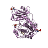
- PDB-1uqy: Xylanase Xyn10B mutant (E262S) from Cellvibrio mixtus in complex ... -
+ Open data
- Basic information
| Entry | Database: PDB / ID: 1uqy | |||||||||
|---|---|---|---|---|---|---|---|---|---|---|
| Title | Xylanase Xyn10B mutant (E262S) from Cellvibrio mixtus in complex with xylopentaose | |||||||||
 Components Components | ENDOXYLANASE | |||||||||
 Keywords Keywords | HYDROLASE / FAMILY 10 / XYLANASE / GLYCOSIDE HYDROLASE / HEMICELLULOSE / XYLAN DEGRADATION | |||||||||
| Function / homology |  Function and homology information Function and homology informationendo-1,4-beta-xylanase activity / endo-1,4-beta-xylanase / xylan catabolic process Similarity search - Function | |||||||||
| Biological species |  CELLVIBRIO MIXTUS (bacteria) CELLVIBRIO MIXTUS (bacteria) | |||||||||
| Method |  X-RAY DIFFRACTION / X-RAY DIFFRACTION /  MOLECULAR REPLACEMENT / Resolution: 1.72 Å MOLECULAR REPLACEMENT / Resolution: 1.72 Å | |||||||||
 Authors Authors | Pell, G. / Taylor, E.J. / Gloster, T.M. / Turkenburg, J.P. / Fontes, C.M.G.A. / Ferreira, L.M.A. / Davies, G.J. / Gilbert, H.J. | |||||||||
 Citation Citation |  Journal: J.Biol.Chem. / Year: 2004 Journal: J.Biol.Chem. / Year: 2004Title: The Mechanisms by which Family 10 Glycoside Hydrolases Bind Decorated Substrates Authors: Pell, G. / Taylor, E.J. / Gloster, T.M. / Turkenburg, J.P. / Fontes, C.M.G.A. / Ferreira, L.M.A. / Nagy, T. / Clark, S. / Davies, G.J. / Gilbert, H.J. | |||||||||
| History |
| |||||||||
| Remark 700 | SHEET DETERMINATION METHOD: DSSP THE SHEETS PRESENTED AS "AA" IN EACH CHAIN ON SHEET RECORDS BELOW ... SHEET DETERMINATION METHOD: DSSP THE SHEETS PRESENTED AS "AA" IN EACH CHAIN ON SHEET RECORDS BELOW IS ACTUALLY AN 10-STRANDED BARREL THIS IS REPRESENTED BY A 11-STRANDED SHEET IN WHICH THE FIRST AND LAST STRANDS ARE IDENTICAL. |
- Structure visualization
| Structure viewer | Molecule:  Molmil Molmil Jmol/JSmol Jmol/JSmol |
|---|
- Downloads & links
- Download
| PDBx/mmCIF format |  1uqy.cif.gz 1uqy.cif.gz | 101 KB | Display |  PDBx/mmCIF format PDBx/mmCIF format |
|---|---|---|---|---|
| PDB format |  pdb1uqy.ent.gz pdb1uqy.ent.gz | 74.5 KB | Display |  PDB format PDB format |
| PDBx/mmJSON format |  1uqy.json.gz 1uqy.json.gz | Tree view |  PDBx/mmJSON format PDBx/mmJSON format | |
| Others |  Other downloads Other downloads |
-Validation report
| Arichive directory |  https://data.pdbj.org/pub/pdb/validation_reports/uq/1uqy https://data.pdbj.org/pub/pdb/validation_reports/uq/1uqy ftp://data.pdbj.org/pub/pdb/validation_reports/uq/1uqy ftp://data.pdbj.org/pub/pdb/validation_reports/uq/1uqy | HTTPS FTP |
|---|
-Related structure data
| Related structure data |  1uqzC  1ur1C 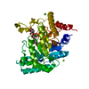 1ur2C  1clxS C: citing same article ( S: Starting model for refinement |
|---|---|
| Similar structure data |
- Links
Links
- Assembly
| Deposited unit | 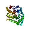
| ||||||||
|---|---|---|---|---|---|---|---|---|---|
| 1 |
| ||||||||
| Unit cell |
|
- Components
| #1: Protein | Mass: 42949.910 Da / Num. of mol.: 1 / Fragment: CATALYTIC DOMAIN, RESIDUES 11-379 / Mutation: YES Source method: isolated from a genetically manipulated source Details: ENGINEERED MUTATION GLU 262 SER IN COORDS / Source: (gene. exp.)  CELLVIBRIO MIXTUS (bacteria) / Plasmid: PET 21A / Production host: CELLVIBRIO MIXTUS (bacteria) / Plasmid: PET 21A / Production host:  | ||
|---|---|---|---|
| #2: Polysaccharide | beta-D-xylopyranose-(1-4)-beta-D-xylopyranose-(1-4)-alpha-D-xylopyranose Source method: isolated from a genetically manipulated source | ||
| #3: Polysaccharide | beta-D-xylopyranose-(1-4)-beta-D-xylopyranose-(1-4)-beta-D-xylopyranose-(1-4)-beta-D-xylopyranose Source method: isolated from a genetically manipulated source | ||
| #4: Chemical | ChemComp-MG / | ||
| #5: Water | ChemComp-HOH / | ||
| Compound details | ENGINEERED| Sequence details | THE SEQUENCE ON THE DATABASE IS INCORRECT AND WILL BE CHANGED BY THE AUTHORS. THESE DISCREPANCIES ...THE SEQUENCE ON THE DATABASE IS INCORRECT AND WILL BE CHANGED BY THE AUTHORS. THESE DISCREPANC | |
-Experimental details
-Experiment
| Experiment | Method:  X-RAY DIFFRACTION / Number of used crystals: 1 X-RAY DIFFRACTION / Number of used crystals: 1 |
|---|
- Sample preparation
| Crystal | Density Matthews: 2 Å3/Da / Density % sol: 36.6 % | ||||||||||||||||||||||||||||||||||||||||||
|---|---|---|---|---|---|---|---|---|---|---|---|---|---|---|---|---|---|---|---|---|---|---|---|---|---|---|---|---|---|---|---|---|---|---|---|---|---|---|---|---|---|---|---|
| Crystal grow | pH: 8 / Details: 0.2M MGCL2 0.1M TRIS HCL PH8, 30% PEG 4K, pH 8.00 | ||||||||||||||||||||||||||||||||||||||||||
| Crystal grow | *PLUS Temperature: 18 ℃ / pH: 8 / Method: vapor diffusion, hanging drop | ||||||||||||||||||||||||||||||||||||||||||
| Components of the solutions | *PLUS
|
-Data collection
| Diffraction | Mean temperature: 120 K |
|---|---|
| Diffraction source | Source:  ROTATING ANODE / Type: RIGAKU RU200 / Wavelength: 1.5418 ROTATING ANODE / Type: RIGAKU RU200 / Wavelength: 1.5418 |
| Detector | Type: MARRESEARCH / Detector: IMAGE PLATE / Date: Feb 15, 2003 / Details: OSMICS |
| Radiation | Monochromator: MULTI-LAYER / Protocol: SINGLE WAVELENGTH / Monochromatic (M) / Laue (L): M / Scattering type: x-ray |
| Radiation wavelength | Wavelength: 1.5418 Å / Relative weight: 1 |
| Reflection | Resolution: 1.72→20 Å / Num. obs: 34539 / % possible obs: 99.6 % / Redundancy: 6.02 % / Rmerge(I) obs: 0.038 / Net I/σ(I): 46 |
| Reflection shell | Resolution: 1.72→1.78 Å / Redundancy: 5.49 % / Rmerge(I) obs: 0.174 / Mean I/σ(I) obs: 10.35 / % possible all: 98.7 |
| Reflection | *PLUS Highest resolution: 1.72 Å / Lowest resolution: 20 Å / Redundancy: 6.02 % / Rmerge(I) obs: 0.038 |
| Reflection shell | *PLUS % possible obs: 98.7 % / Rmerge(I) obs: 0.174 / Mean I/σ(I) obs: 10.35 |
- Processing
| Software |
| ||||||||||||||||||||||||||||||||||||||||||||||||||||||||||||||||||||||||||||||||||||||||||||||||||||||||||||||||||||||||||||||||||||||||||||||||||||||||||||||||||||||||||||||||||||||
|---|---|---|---|---|---|---|---|---|---|---|---|---|---|---|---|---|---|---|---|---|---|---|---|---|---|---|---|---|---|---|---|---|---|---|---|---|---|---|---|---|---|---|---|---|---|---|---|---|---|---|---|---|---|---|---|---|---|---|---|---|---|---|---|---|---|---|---|---|---|---|---|---|---|---|---|---|---|---|---|---|---|---|---|---|---|---|---|---|---|---|---|---|---|---|---|---|---|---|---|---|---|---|---|---|---|---|---|---|---|---|---|---|---|---|---|---|---|---|---|---|---|---|---|---|---|---|---|---|---|---|---|---|---|---|---|---|---|---|---|---|---|---|---|---|---|---|---|---|---|---|---|---|---|---|---|---|---|---|---|---|---|---|---|---|---|---|---|---|---|---|---|---|---|---|---|---|---|---|---|---|---|---|---|
| Refinement | Method to determine structure:  MOLECULAR REPLACEMENT MOLECULAR REPLACEMENTStarting model: PDB ENTRY 1CLX Resolution: 1.72→56.8 Å / Cor.coef. Fo:Fc: 0.97 / Cor.coef. Fo:Fc free: 0.954 / SU B: 1.774 / SU ML: 0.06 / Cross valid method: THROUGHOUT / ESU R: 0.1 / ESU R Free: 0.103 / Stereochemistry target values: MAXIMUM LIKELIHOOD Details: HYDROGENS HAVE BEEN ADDED IN THE RIDING POSITIONS. THIS ENTRY HAS SOME ATOMS WHICH HAVE BEEN REFINED WITH AN OCCUPANCY OF 0.00
| ||||||||||||||||||||||||||||||||||||||||||||||||||||||||||||||||||||||||||||||||||||||||||||||||||||||||||||||||||||||||||||||||||||||||||||||||||||||||||||||||||||||||||||||||||||||
| Solvent computation | Ion probe radii: 0.8 Å / Shrinkage radii: 0.8 Å / VDW probe radii: 1.2 Å / Solvent model: BABINET MODEL WITH MASK | ||||||||||||||||||||||||||||||||||||||||||||||||||||||||||||||||||||||||||||||||||||||||||||||||||||||||||||||||||||||||||||||||||||||||||||||||||||||||||||||||||||||||||||||||||||||
| Displacement parameters | Biso mean: 12.79 Å2
| ||||||||||||||||||||||||||||||||||||||||||||||||||||||||||||||||||||||||||||||||||||||||||||||||||||||||||||||||||||||||||||||||||||||||||||||||||||||||||||||||||||||||||||||||||||||
| Refinement step | Cycle: LAST / Resolution: 1.72→56.8 Å
| ||||||||||||||||||||||||||||||||||||||||||||||||||||||||||||||||||||||||||||||||||||||||||||||||||||||||||||||||||||||||||||||||||||||||||||||||||||||||||||||||||||||||||||||||||||||
| Refine LS restraints |
|
 Movie
Movie Controller






 PDBj
PDBj


