 Movie
Movie Controller

[English] 日本語
 Yorodumi
- PDB-1up9: REDUCED STRUCTURE OF CYTOCHROME C3 FROM DESULFOVIBRIO DESULFURICA... -
+ Open data

- Basic information
| Entry | Database: PDB / ID: 1up9 | ||||||
|---|---|---|---|---|---|---|---|
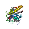
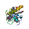
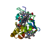
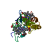
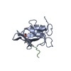
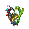
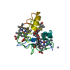
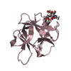
| Title | REDUCED STRUCTURE OF CYTOCHROME C3 FROM DESULFOVIBRIO DESULFURICANS ATCC 27774 AT PH 7.6 | ||||||
 Components Components | CYTOCHROME C3 | ||||||
 Keywords Keywords | ELECTRON TRANSPORT / TETRAHEME CYTOCHROME C | ||||||
| Function / homology |  Function and homology information Function and homology information | ||||||
| Biological species |  DESULFOVIBRIO DESULFURICANS (bacteria) DESULFOVIBRIO DESULFURICANS (bacteria) | ||||||
| Method |  X-RAY DIFFRACTION / SYNCHROTRON / MOLECULAR REPLACEMENT / Resolution: 1.35 Å X-RAY DIFFRACTION / SYNCHROTRON / MOLECULAR REPLACEMENT / Resolution: 1.35 Å | ||||||
 Authors Authors | Bento, I. / Matias, P.M. / Baptista, A.M. / Da Costa, P.N. / Van Dongen, W.M.A.M. / Saraiva, L.M. / Schneider, T.R. / Soares, C.M. / Carrondo, M.A. | ||||||
 Citation Citation | Journal: Proteins / Year: 2004 Title: Molecular Basis for Redox-Bohr and Cooperative Effects in Cytochrome C3 from Desulfovibrio Desulfuricans Atcc 27774: Crystallographic and Modeling Studies of Oxidized and Reduced High- ...Title: Molecular Basis for Redox-Bohr and Cooperative Effects in Cytochrome C3 from Desulfovibrio Desulfuricans Atcc 27774: Crystallographic and Modeling Studies of Oxidized and Reduced High-Resolution Structures at Ph 7.6 Authors: Bento, I. / Matias, P.M. / Baptista, A.M. / Da Costa, P.N. / Van Dongen, W.M.A.M. / Saraiva, L.M. / Schneider, T.R. / Soares, C.M. / Carrondo, M.A. #1: Journal: J.Biol.Chem. / Year: 2001Title: Conformational Component in the Coupled Transfer of Multiple Electrons and Protons in a Monomeric Tetraheme Cytochrome Authors: Louro, R.O. / Bento, I. / Matias, P.M. / Catarino, T. / Baptista, A.M. / Soares, C.M. / Carrondo, M.A. / Turner, D.L. / Xavier, A.V. #2: Journal: Inorg.Chim.Acta. / Year: 1998Title: Refinement of the Three-Dimensional Structures of Cytochrome C3 from Desulfovibrio Vulgaris Hildenborough at 1.67 A Resolution and from Desulfovibrio Desulfuricans Atcc 27774 at 1.6 A Resolution Authors: Simoes, P. / Matias, P.M. / Morais, J. / Wilson, K. / Dauter, Z. / Carrondo, M.A. #3: Journal: Biochemistry / Year: 1995 Title: Structure of the Tetraheme Cytochrome from Desulfovibrio Desulfuricans Atcc 27774: X-Ray Diffraction and Electron Paramagnetic Resonance Studies Authors: Morais, J. / Palma, P.N. / Frazao, C. / Caldeira, J. / Legall, J. / Moura, I. / Moura, J.J. / Carrondo, M.A. | ||||||
| History |
|
- Structure visualization
| Structure viewer | Molecule: MolmilJmol/JSmol |
|---|
- Downloads & links
-Download
| PDBx/mmCIF format | 1up9.cif.gz | 72.7 KB | Display | PDBx/mmCIF format |
|---|---|---|---|---|
| PDB format | pdb1up9.ent.gz | 55.2 KB | Display | PDB format |
| PDBx/mmJSON format | 1up9.json.gz | Tree view | PDBx/mmJSON format | |
| Others |  Other downloads Other downloads |
-Validation report
| Arichive directory | https://data.pdbj.org/pub/pdb/validation_reports/up/1up9ftp://data.pdbj.org/pub/pdb/validation_reports/up/1up9 | HTTPS FTP |
|---|
-Related structure data
| Related structure data |  1updC 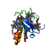 3cyrS C: citing same article ( S: Starting model for refinement |
|---|---|
| Similar structure data |


-Links
 PDBj
PDBj


- Assembly
| Deposited unit | 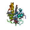
| |||||||||||||||
|---|---|---|---|---|---|---|---|---|---|---|---|---|---|---|---|---|
| 1 |
| |||||||||||||||
| Unit cell |
| |||||||||||||||
| Components on special symmetry positions |
|
-Components
| #1: Protein | Mass: 11618.471 Da / Num. of mol.: 1 / Source method: isolated from a natural source / Source: (natural) DESULFOVIBRIO DESULFURICANS (bacteria) / References: UniProt: Q9L915 | ||||||
|---|---|---|---|---|---|---|---|
| #2: Chemical | ChemComp-HEC /   Mass: 618.503 Da / Num. of mol.: 4 / Source method: obtained synthetically / Formula: C34H34FeN4O4 Mass: 618.503 Da / Num. of mol.: 4 / Source method: obtained synthetically / Formula: C34H34FeN4O4#3: Chemical | ChemComp-SO4 / |   Mass: 96.063 Da / Num. of mol.: 1 / Source method: obtained synthetically / Formula: SO4 Mass: 96.063 Da / Num. of mol.: 1 / Source method: obtained synthetically / Formula: SO4#4: Water | ChemComp-HOH / |  Mass: 18.015 Da / Num. of mol.: 144 / Source method: isolated from a natural source / Formula: H2O Mass: 18.015 Da / Num. of mol.: 144 / Source method: isolated from a natural source / Formula: H2OHas protein modification | Y | |
-Experimental details
-Experiment
| Experiment | Method: X-RAY DIFFRACTION / Number of used crystals: 1 |
|---|
- Sample preparation
| Crystal | Density Matthews: 1.86 Å3/Da / Density % sol: 34 % |
|---|---|
| Crystal grow | pH: 7.6 / Details: pH 7.60 |
-Data collection
| Diffraction | Mean temperature: 110 K |
|---|---|
| Diffraction source | Source: SYNCHROTRON / Site: ESRF  / Beamline: ID14-3 / Wavelength: 0.931 / Beamline: ID14-3 / Wavelength: 0.931 |
| Detector | Type: MARRESEARCH / Detector: CCD / Details: TOROIDAL MIRROR |
| Radiation | Monochromator: DIAMOND (III) CRYSTAL / Protocol: SINGLE WAVELENGTH / Monochromatic (M) / Laue (L): M / Scattering type: x-ray |
| Radiation wavelength | Wavelength: 0.931 Å / Relative weight: 1 |
| Reflection | Resolution: 1.35→30 Å / Num. obs: 26954 / % possible obs: 99.5 % / Observed criterion σ(I): 0 / Redundancy: 4.14 % / Rmerge(I) obs: 0.033 |
| Reflection shell | Resolution: 1.35→1.38 Å / Rmerge(I) obs: 0.183 / % possible all: 96.8 |
- Processing
| Software |
| |||||||||||||||||||||||||||||||||
|---|---|---|---|---|---|---|---|---|---|---|---|---|---|---|---|---|---|---|---|---|---|---|---|---|---|---|---|---|---|---|---|---|---|---|
| Refinement | Method to determine structure: MOLECULAR REPLACEMENT Starting model: PDB ENTRY 3CYR Resolution: 1.35→30 Å / Num. parameters: 10545 / Num. restraintsaints: 13944 / Cross valid method: FREE R-VALUE / σ(F): 0 / Stereochemistry target values: ENGH AND HUBER
| |||||||||||||||||||||||||||||||||
| Solvent computation | Solvent model: MOEWS & KRETSINGER | |||||||||||||||||||||||||||||||||
| Refine analyze | Num. disordered residues: 14 / Occupancy sum hydrogen: 0 / Occupancy sum non hydrogen: 1107.54 | |||||||||||||||||||||||||||||||||
| Refinement step | Cycle: LAST / Resolution: 1.35→30 Å
| |||||||||||||||||||||||||||||||||
| Refine LS restraints |
|
