 ムービー
ムービー コントローラー


+ データを開く

- 基本情報
基本情報
| 登録情報 | データベース: PDB / ID: 1gmb | ||||||
|---|---|---|---|---|---|---|---|
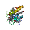
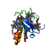
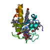
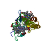
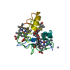
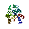
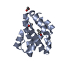
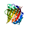
| タイトル | Reduced structure of CYTOCHROME C3 FROM DESULFOVIBRIO DESULFURICANS ATCC 27774 at pH 7.6 | ||||||
 要素 要素 | CYTOCHROME C3 | ||||||
 キーワード キーワード | ELECTRON TRANSPORT / CYTOCHROME | ||||||
| 機能・相同性 |  機能・相同性情報 機能・相同性情報 | ||||||
| 生物種 |  DESULFOVIBRIO DESULFURICANS (バクテリア) DESULFOVIBRIO DESULFURICANS (バクテリア) | ||||||
| 手法 |  X線回折 / 分子置換 / 解像度: 2 Å X線回折 / 分子置換 / 解像度: 2 Å | ||||||
 データ登録者 データ登録者 | Bento, I. / Louro, R. / Matias, P.M. / Catarino, T. / Baptista, A.M. / Soares, C.M. / Carrondo, M.A. / Turner, D.L. / Xavier, A.V. | ||||||
 引用 引用 | ジャーナル: J.Biol.Chem. / 年: 2001 タイトル: Conformational Component in the Coupled Transfer of Multiple Electrons and Protons in a Monomeric Tetraheme Cytochrome 著者: Louro, R. / Bento, I. / Matias, P.M. / Catarino, T. / Baptista, A.M. / Soares, C.M. / Carrondo, M.A. / Turner, D.L. / Xavier, A.V. #1: ジャーナル: Inorg.Chim.Acta / 年: 1998タイトル: Refinement of the Three-Dimensional Structures of Cytochrome C3 from Desulfovibrio Vulgaris Hildenborough at 1.67 Angstroms Resolution and from Desulfovibrio Desulfuricans Atcc 27774 at ...タイトル: Refinement of the Three-Dimensional Structures of Cytochrome C3 from Desulfovibrio Vulgaris Hildenborough at 1.67 Angstroms Resolution and from Desulfovibrio Desulfuricans Atcc 27774 at 1.6 Angstrom S Resolution 著者: Simoes, P. / Matias, P.M. / Morais, J. / Wilson, K. / Dauter, Z. / Carrondo, M.A. #2: ジャーナル: Biochemistry / 年: 1995 タイトル: Structure of the Tetraheme Cytochrome from Desulfovibrio Desulfuricans Atcc 27774: X-Ray Diffraction and Electron Paramagnetic Resonance Studies 著者: Morais, J. / Palma, P.N. / Frazao, C. / Caldeira, J. / Legall, J. / Moura, I. / Moura, J.J. / Carrondo, M.A. | ||||||
| 履歴 |
|
- 構造の表示
構造の表示
| 構造ビューア | 分子: MolmilJmol/JSmol |
|---|
- ダウンロードとリンク
-ダウンロード
| PDBx/mmCIF形式 | 1gmb.cif.gz | 40.7 KB | 表示 | PDBx/mmCIF形式 |
|---|---|---|---|---|
| PDB形式 | pdb1gmb.ent.gz | 28.7 KB | 表示 | PDB形式 |
| PDBx/mmJSON形式 | 1gmb.json.gz | ツリー表示 | PDBx/mmJSON形式 | |
| その他 |  その他のダウンロード その他のダウンロード |
-検証レポート
| アーカイブディレクトリ | https://data.pdbj.org/pub/pdb/validation_reports/gm/1gmbftp://data.pdbj.org/pub/pdb/validation_reports/gm/1gmb | HTTPS FTP |
|---|
-関連構造データ


-リンク
 PDBj
PDBj


- 集合体
集合体
| 登録構造単位 | 
| ||||||||
|---|---|---|---|---|---|---|---|---|---|
| 1 |
| ||||||||
| 単位格子 |
|
-要素
| #1: タンパク質 | 分子量: 11618.471 Da / 分子数: 1 / 由来タイプ: 天然 由来: (天然) DESULFOVIBRIO DESULFURICANS (バクテリア)参照: UniProt: Q9L915 | ||||||
|---|---|---|---|---|---|---|---|
| #2: 化合物 | ChemComp-HEC /   分子量: 618.503 Da / 分子数: 4 / 由来タイプ: 合成 / 式: C34H34FeN4O4 分子量: 618.503 Da / 分子数: 4 / 由来タイプ: 合成 / 式: C34H34FeN4O4#3: 化合物 |   分子量: 96.063 Da / 分子数: 2 / 由来タイプ: 合成 / 式: SO4 分子量: 96.063 Da / 分子数: 2 / 由来タイプ: 合成 / 式: SO4#4: 水 | ChemComp-HOH / |  分子量: 18.015 Da / 分子数: 97 / 由来タイプ: 天然 / 式: H2O 分子量: 18.015 Da / 分子数: 97 / 由来タイプ: 天然 / 式: H2OHas protein modification | Y | |
-実験情報
-実験
| 実験 | 手法: X線回折 / 使用した結晶の数: 1 |
|---|
- 試料調製
試料調製
| 結晶 | マシュー密度: 2.5 Å3/Da / 溶媒含有率: 50.71 % |
|---|---|
| 結晶化 | pH: 7.6 / 詳細: pH 7.60 |
| 結晶化 | *PLUS 手法: other / 詳細: Morais, J., (1995) Biochemistry, 34, 12830. |
-データ収集
| 回折 | 平均測定温度: 110 K |
|---|---|
| 放射光源 | 由来: 回転陽極 / タイプ: ENRAF-NONIUS FR571 / 波長: 1.5418 |
| 検出器 | タイプ: MARRESEARCH / 検出器: IMAGE PLATE |
| 放射 | モノクロメーター: GRAPHITE / プロトコル: SINGLE WAVELENGTH / 単色(M)・ラウエ(L): M / 散乱光タイプ: x-ray |
| 放射波長 | 波長: 1.5418 Å / 相対比: 1 |
| 反射 | 解像度: 2→24 Å / Num. obs: 8507 / % possible obs: 99.3 % / Observed criterion σ(I): 0 / 冗長度: 6 % / Rmerge(I) obs: 0.048 |
| 反射 シェル | 解像度: 2→2.07 Å / Rmerge(I) obs: 0.186 / % possible all: 99.8 |
| 反射 | *PLUS 最高解像度: 2 Å / Num. measured all: 54926 |
| 反射 シェル | *PLUS % possible obs: 99.8 % |
- 解析
解析
| ソフトウェア |
| ||||||||||||||||||||||||||||||||||||||||||||||||||||||||||||
|---|---|---|---|---|---|---|---|---|---|---|---|---|---|---|---|---|---|---|---|---|---|---|---|---|---|---|---|---|---|---|---|---|---|---|---|---|---|---|---|---|---|---|---|---|---|---|---|---|---|---|---|---|---|---|---|---|---|---|---|---|---|
| 精密化 | 構造決定の手法: 分子置換 開始モデル: PDB ENTRY 3CYR 解像度: 2→24 Å / 交差検証法: FREE R-VALUE / σ(F): 2
| ||||||||||||||||||||||||||||||||||||||||||||||||||||||||||||
| 精密化ステップ | サイクル: LAST / 解像度: 2→24 Å
| ||||||||||||||||||||||||||||||||||||||||||||||||||||||||||||
| 拘束条件 |
| ||||||||||||||||||||||||||||||||||||||||||||||||||||||||||||
| 精密化 | *PLUS 最高解像度: 2 Å | ||||||||||||||||||||||||||||||||||||||||||||||||||||||||||||
| 溶媒の処理 | *PLUS | ||||||||||||||||||||||||||||||||||||||||||||||||||||||||||||
| 原子変位パラメータ | *PLUS |
