 Movie
Movie Controller

[English] 日本語
 Yorodumi
- PDB-1unn: Complex of beta-clamp processivity factor and little finger domai... -
+ Open data

- Basic information
| Entry | Database: PDB / ID: 1unn | ||||||
|---|---|---|---|---|---|---|---|
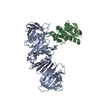
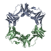
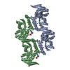
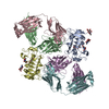
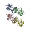
| Title | Complex of beta-clamp processivity factor and little finger domain of PolIV | ||||||
 Components Components |
| ||||||
 Keywords Keywords | BETA-CLAMP / POL IV / TRANSLESION / TRANSFERASE / DNA-DIRECTED DNA POLYMERASE / DNA REPLICATION | ||||||
| Function / homology |  Function and homology information Function and homology informationHda-beta clamp complex / bacterial-type DNA replication / replication inhibiting complex / DNA polymerase III complex / SOS response / replisome / regulation of DNA-templated DNA replication initiation / error-free translesion synthesis / DNA strand elongation involved in DNA replication / DNA synthesis involved in DNA repair ...Hda-beta clamp complex / bacterial-type DNA replication / replication inhibiting complex / DNA polymerase III complex / SOS response / replisome / regulation of DNA-templated DNA replication initiation / error-free translesion synthesis / DNA strand elongation involved in DNA replication / DNA synthesis involved in DNA repair / error-prone translesion synthesis / 3'-5' exonuclease activity / negative regulation of DNA-templated DNA replication initiation / DNA-templated DNA replication / DNA-directed DNA polymerase / damaged DNA binding / DNA-directed DNA polymerase activity / DNA replication / DNA damage response / magnesium ion binding / protein homodimerization activity / DNA binding / identical protein binding / cytoplasm / cytosol Similarity search - Function | ||||||
| Biological species |  | ||||||
| Method |  X-RAY DIFFRACTION / SYNCHROTRON / MOLECULAR REPLACEMENT / Resolution: 1.9 Å X-RAY DIFFRACTION / SYNCHROTRON / MOLECULAR REPLACEMENT / Resolution: 1.9 Å | ||||||
 Authors Authors | Bunting, K.A. / Roe, S.M. / Pearl, L.H. | ||||||
 Citation Citation | Journal: Embo J. / Year: 2003 Title: Structural Basis for Recruitment of Translesion DNA Polymerase Pol Iv/Dinb to the Beta-Clamp Authors: Bunting, K.A. / Roe, S.M. / Pearl, L.H. | ||||||
| History |
| ||||||
| Remark 700 | SHEET THE SHEET STRUCTURE OF THIS MOLECULE IS BIFURCATED. IN ORDER TO REPRESENT THIS FEATURE IN ... SHEET THE SHEET STRUCTURE OF THIS MOLECULE IS BIFURCATED. IN ORDER TO REPRESENT THIS FEATURE IN THE SHEET RECORDS BELOW, TWO SHEETS ARE DEFINED. |
- Structure visualization
| Structure viewer | Molecule: MolmilJmol/JSmol |
|---|
- Downloads & links
-Download
| PDBx/mmCIF format | 1unn.cif.gz | 219.2 KB | Display | PDBx/mmCIF format |
|---|---|---|---|---|
| PDB format | pdb1unn.ent.gz | 175.9 KB | Display | PDB format |
| PDBx/mmJSON format | 1unn.json.gz | Tree view | PDBx/mmJSON format | |
| Others |  Other downloads Other downloads |
-Validation report
| Arichive directory | https://data.pdbj.org/pub/pdb/validation_reports/un/1unnftp://data.pdbj.org/pub/pdb/validation_reports/un/1unn | HTTPS FTP |
|---|
-Related structure data
| Related structure data |  2polS S: Starting model for refinement |
|---|---|
| Similar structure data |


-Links
 PDBj
PDBj


- Assembly
| Deposited unit | 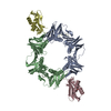
| ||||||||
|---|---|---|---|---|---|---|---|---|---|
| 1 |
| ||||||||
| Unit cell |
| ||||||||
| Components on special symmetry positions |
|
-Components
| #1: Protein | Mass: 40630.508 Da / Num. of mol.: 2 Source method: isolated from a genetically manipulated source Source: (gene. exp.) References: UniProt: P00583, UniProt: P0A988*PLUS, DNA-directed DNA polymerase #2: Protein | Mass: 13556.599 Da / Num. of mol.: 2 / Fragment: LITTLE FINGER, RESIDUES 243-351 Source method: isolated from a genetically manipulated source Source: (gene. exp.) #3: Chemical |   Mass: 96.063 Da / Num. of mol.: 2 / Source method: obtained synthetically / Formula: SO4 Mass: 96.063 Da / Num. of mol.: 2 / Source method: obtained synthetically / Formula: SO4#4: Water | ChemComp-HOH / |  Mass: 18.015 Da / Num. of mol.: 947 / Source method: isolated from a natural source / Formula: H2O Mass: 18.015 Da / Num. of mol.: 947 / Source method: isolated from a natural source / Formula: H2O |
|---|
-Experimental details
-Experiment
| Experiment | Method: X-RAY DIFFRACTION / Number of used crystals: 1 |
|---|
- Sample preparation
| Crystal | Density Matthews: 2.4 Å3/Da / Density % sol: 48.28 % | ||||||||||||||||||||||||
|---|---|---|---|---|---|---|---|---|---|---|---|---|---|---|---|---|---|---|---|---|---|---|---|---|---|
| Crystal grow | Temperature: 287 K / Method: vapor diffusion, hanging drop / pH: 4.6 Details: CRYSTALS WERE OBTAINED USING HANGING DROP METHOD AT 14C. WELL CONSISTED OF 1.2M AMMONIUM SULPHATE, 50MM SODIUM ACETATE PH 4.6. 2UL DROP CONSISTED OF 1UL WELL SOLUTION PLUS 1UL PROTEIN ...Details: CRYSTALS WERE OBTAINED USING HANGING DROP METHOD AT 14C. WELL CONSISTED OF 1.2M AMMONIUM SULPHATE, 50MM SODIUM ACETATE PH 4.6. 2UL DROP CONSISTED OF 1UL WELL SOLUTION PLUS 1UL PROTEIN SOLUTION (50MM HEPES PH7.0, 2MM MAGNESIUM CHLORIDE, 200MM SODIUM CHLORIDE AND PROTEIN AT 6MG/ML). | ||||||||||||||||||||||||
| Crystal grow | *PLUS Temperature: 14 ℃ / pH: 4.6 / Method: vapor diffusion, hanging drop | ||||||||||||||||||||||||
| Components of the solutions | *PLUS
|
-Data collection
| Diffraction | Mean temperature: 100 K |
|---|---|
| Diffraction source | Source: SYNCHROTRON / Site: ESRF  / Beamline: ID14-1 / Wavelength: 0.934 / Beamline: ID14-1 / Wavelength: 0.934 |
| Detector | Type: ADSC CCD / Detector: CCD / Date: Nov 15, 2002 |
| Radiation | Protocol: SINGLE WAVELENGTH / Monochromatic (M) / Laue (L): M / Scattering type: x-ray |
| Radiation wavelength | Wavelength: 0.934 Å / Relative weight: 1 |
| Reflection | Resolution: 1.9→100 Å / Num. obs: 90134 / % possible obs: 96.5 % / Redundancy: 4.9 % / Rmerge(I) obs: 0.06 / Net I/σ(I): 8.3 |
| Reflection shell | Resolution: 1.9→2.01 Å / Redundancy: 2.7 % / Rmerge(I) obs: 0.201 / Mean I/σ(I) obs: 3.5 / % possible all: 87.4 |
| Reflection | *PLUS Highest resolution: 1.9 Å / Lowest resolution: 62.1 Å / Redundancy: 4.9 % / Rmerge(I) obs: 0.06 |
| Reflection shell | *PLUS % possible obs: 87.4 % / Redundancy: 2.7 % / Num. unique obs: 12565 / Rmerge(I) obs: 0.201 / Mean I/σ(I) obs: 3.5 |
- Processing
| Software |
| ||||||||||||||||||||||||||||||||||||||||||||||||||||||||||||||||||||||||||||||||||||||||||||||||||||||||||||||||||||||||||||||||||||||||||||||||||||||||||||||||||||||||||||||||||||||
|---|---|---|---|---|---|---|---|---|---|---|---|---|---|---|---|---|---|---|---|---|---|---|---|---|---|---|---|---|---|---|---|---|---|---|---|---|---|---|---|---|---|---|---|---|---|---|---|---|---|---|---|---|---|---|---|---|---|---|---|---|---|---|---|---|---|---|---|---|---|---|---|---|---|---|---|---|---|---|---|---|---|---|---|---|---|---|---|---|---|---|---|---|---|---|---|---|---|---|---|---|---|---|---|---|---|---|---|---|---|---|---|---|---|---|---|---|---|---|---|---|---|---|---|---|---|---|---|---|---|---|---|---|---|---|---|---|---|---|---|---|---|---|---|---|---|---|---|---|---|---|---|---|---|---|---|---|---|---|---|---|---|---|---|---|---|---|---|---|---|---|---|---|---|---|---|---|---|---|---|---|---|---|---|
| Refinement | Method to determine structure: MOLECULAR REPLACEMENT Starting model: PDB ENTRY 2POL Resolution: 1.9→62.1 Å / Cor.coef. Fo:Fc: 0.951 / Cor.coef. Fo:Fc free: 0.914 / SU B: 2.981 / SU ML: 0.09 / TLS residual ADP flag: LIKELY RESIDUAL / Cross valid method: THROUGHOUT / ESU R: 0.146 / ESU R Free: 0.15 / Stereochemistry target values: MAXIMUM LIKELIHOOD
| ||||||||||||||||||||||||||||||||||||||||||||||||||||||||||||||||||||||||||||||||||||||||||||||||||||||||||||||||||||||||||||||||||||||||||||||||||||||||||||||||||||||||||||||||||||||
| Solvent computation | Ion probe radii: 0.8 Å / Shrinkage radii: 0.8 Å / VDW probe radii: 1.4 Å / Solvent model: BABINET MODEL PLUS MASK | ||||||||||||||||||||||||||||||||||||||||||||||||||||||||||||||||||||||||||||||||||||||||||||||||||||||||||||||||||||||||||||||||||||||||||||||||||||||||||||||||||||||||||||||||||||||
| Displacement parameters | Biso mean: 21.92 Å2
| ||||||||||||||||||||||||||||||||||||||||||||||||||||||||||||||||||||||||||||||||||||||||||||||||||||||||||||||||||||||||||||||||||||||||||||||||||||||||||||||||||||||||||||||||||||||
| Refinement step | Cycle: LAST / Resolution: 1.9→62.1 Å
| ||||||||||||||||||||||||||||||||||||||||||||||||||||||||||||||||||||||||||||||||||||||||||||||||||||||||||||||||||||||||||||||||||||||||||||||||||||||||||||||||||||||||||||||||||||||
| Refine LS restraints |
|