 Movie
Movie Controller

[English] 日本語
 Yorodumi
- PDB-7d85: Crystal structure of anti-ErbB3 Fab ISU104 in complex with human ... -
+ Open data

- Basic information
| Entry | Database: PDB / ID: 7d85 | ||||||
|---|---|---|---|---|---|---|---|
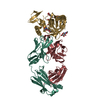
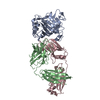
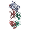
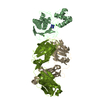
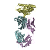
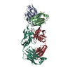
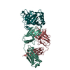
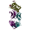
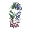
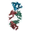
| Title | Crystal structure of anti-ErbB3 Fab ISU104 in complex with human ErbB3 extracellular domain 3 | ||||||
 Components Components |
| ||||||
 Keywords Keywords | IMMUNE SYSTEM / Transferase/Immune System | ||||||
| Function / homology |  Function and homology information Function and homology informationpositive regulation of cardiac muscle tissue development / neuregulin binding / cranial nerve development / Schwann cell differentiation / neuregulin receptor activity / negative regulation of secretion / endocardial cushion development / ERBB3:ERBB2 complex / GRB7 events in ERBB2 signaling / positive regulation of calcineurin-NFAT signaling cascade ...positive regulation of cardiac muscle tissue development / neuregulin binding / cranial nerve development / Schwann cell differentiation / neuregulin receptor activity / negative regulation of secretion / endocardial cushion development / ERBB3:ERBB2 complex / GRB7 events in ERBB2 signaling / positive regulation of calcineurin-NFAT signaling cascade / peripheral nervous system development / negative regulation of cell adhesion / ErbB-3 class receptor binding / negative regulation of motor neuron apoptotic process / motor neuron apoptotic process / ERBB2 Activates PTK6 Signaling / growth factor binding / ERBB2-ERBB3 signaling pathway / ERBB2 Regulates Cell Motility / protein tyrosine kinase activator activity / Signaling by ERBB4 / PI3K events in ERBB2 signaling / lateral plasma membrane / negative regulation of signal transduction / Schwann cell development / extrinsic apoptotic signaling pathway in absence of ligand / Signaling by ERBB2 / myelination / SHC1 events in ERBB2 signaling / cell surface receptor protein tyrosine kinase signaling pathway / basal plasma membrane / positive regulation of epithelial cell proliferation / Downregulation of ERBB2:ERBB3 signaling / wound healing / phosphatidylinositol 3-kinase/protein kinase B signal transduction / Signaling by ERBB2 TMD/JMD mutants / receptor protein-tyrosine kinase / Signaling by ERBB2 KD Mutants / epidermal growth factor receptor signaling pathway / Downregulation of ERBB2 signaling / Constitutive Signaling by Aberrant PI3K in Cancer / neuron differentiation / transmembrane signaling receptor activity / PIP3 activates AKT signaling / regulation of cell population proliferation / heart development / PI5P, PP2A and IER3 Regulate PI3K/AKT Signaling / RAF/MAP kinase cascade / neuron apoptotic process / basolateral plasma membrane / negative regulation of neuron apoptotic process / protein kinase activity / positive regulation of MAPK cascade / positive regulation of phosphatidylinositol 3-kinase/protein kinase B signal transduction / signaling receptor complex / apical plasma membrane / protein heterodimerization activity / ubiquitin protein ligase binding / positive regulation of gene expression / negative regulation of apoptotic process / signal transduction / : / ATP binding / identical protein binding / plasma membrane Similarity search - Function | ||||||
| Biological species |  Homo sapiens (human) Homo sapiens (human) | ||||||
| Method |  X-RAY DIFFRACTION / SYNCHROTRON / SAD / Resolution: 2.5 Å X-RAY DIFFRACTION / SYNCHROTRON / SAD / Resolution: 2.5 Å | ||||||
 Authors Authors | Yoo, Y. / Cho, H.S. | ||||||
 Citation Citation | Journal: Mol.Cancer Ther. / Year: 2021 Title: A Novel Therapeutic Anti-ErbB3, ISU104 Exhibits Potent Antitumorigenic Activity by Inhibiting Ligand Binding and ErbB3 Heterodimerization. Authors: Hong, M. / Yoo, Y. / Kim, M. / Kim, J.Y. / Cha, J.S. / Choi, M.K. / Kim, U. / Kim, K. / Sohn, Y. / Bae, D. / Cho, H.S. / Hong, S.B. | ||||||
| History |
|
- Structure visualization
| Structure viewer | Molecule: MolmilJmol/JSmol |
|---|
- Downloads & links
-Download
| PDBx/mmCIF format | 7d85.cif.gz | 265.7 KB | Display | PDBx/mmCIF format |
|---|---|---|---|---|
| PDB format | pdb7d85.ent.gz | 194.8 KB | Display | PDB format |
| PDBx/mmJSON format | 7d85.json.gz | Tree view | PDBx/mmJSON format | |
| Others |  Other downloads Other downloads |
-Validation report
| Arichive directory | https://data.pdbj.org/pub/pdb/validation_reports/d8/7d85ftp://data.pdbj.org/pub/pdb/validation_reports/d8/7d85 | HTTPS FTP |
|---|
-Related structure data
| Similar structure data |
|---|

-Links
 PDBj
PDBj


- Assembly
| Deposited unit | 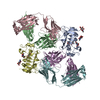
| ||||||||||||
|---|---|---|---|---|---|---|---|---|---|---|---|---|---|
| 1 | 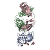
| ||||||||||||
| 2 | 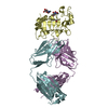
| ||||||||||||
| Unit cell |
|
-Components
| #1: Protein | Mass: 21490.555 Da / Num. of mol.: 2 / Fragment: extracellular domain 3 Source method: isolated from a genetically manipulated source Source: (gene. exp.) Homo sapiens (human) / Gene: ERBB3, HER3 / Production host:   Spodoptera frugiperda (fall armyworm) Spodoptera frugiperda (fall armyworm)References: UniProt: P21860, receptor protein-tyrosine kinase #2: Antibody | Mass: 24252.021 Da / Num. of mol.: 2 Source method: isolated from a genetically manipulated source Source: (gene. exp.) Homo sapiens (human) / Production host:   Cricetulus griseus (Chinese hamster) Cricetulus griseus (Chinese hamster)#3: Antibody | Mass: 22663.936 Da / Num. of mol.: 2 Source method: isolated from a genetically manipulated source Source: (gene. exp.) Homo sapiens (human) / Production host: Cricetulus griseus (Chinese hamster)#4: Sugar | ChemComp-NAG /   Type: D-saccharide, beta linking / Mass: 221.208 Da / Num. of mol.: 7 / Source method: obtained synthetically / Formula: C8H15NO6 Type: D-saccharide, beta linking / Mass: 221.208 Da / Num. of mol.: 7 / Source method: obtained synthetically / Formula: C8H15NO6#5: Water | ChemComp-HOH / |  Mass: 18.015 Da / Num. of mol.: 39 / Source method: isolated from a natural source / Formula: H2O Mass: 18.015 Da / Num. of mol.: 39 / Source method: isolated from a natural source / Formula: H2OHas ligand of interest | N | Has protein modification | Y | |
|---|
-Experimental details
-Experiment
| Experiment | Method: X-RAY DIFFRACTION / Number of used crystals: 1 |
|---|
- Sample preparation
| Crystal | Density Matthews: 2.26 Å3/Da / Density % sol: 45.61 % |
|---|---|
| Crystal grow | Temperature: 298 K / Method: vapor diffusion, sitting drop / Details: 0.2M Sodium acetate, 20%(w/v) PEG 3350 |
-Data collection
| Diffraction | Mean temperature: 100 K / Serial crystal experiment: N |
|---|---|
| Diffraction source | Source: SYNCHROTRON / Site: PAL/PLS  / Beamline: 11C / Wavelength: 0.98 Å / Beamline: 11C / Wavelength: 0.98 Å |
| Detector | Type: DECTRIS PILATUS 6M / Detector: PIXEL / Date: Mar 26, 2020 |
| Radiation | Protocol: SINGLE WAVELENGTH / Monochromatic (M) / Laue (L): M / Scattering type: x-ray |
| Radiation wavelength | Wavelength: 0.98 Å / Relative weight: 1 |
| Reflection | Resolution: 2.5→46.63 Å / Num. obs: 42145 / % possible obs: 99.2 % / Redundancy: 2 % / Biso Wilson estimate: 36.1 Å2 / CC1/2: 0.987 / CC star: 0.997 / Rmerge(I) obs: 0.09686 / Net I/σ(I): 6.56 |
| Reflection shell | Resolution: 2.5→2.589 Å / Redundancy: 2 % / Rmerge(I) obs: 0.5366 / Mean I/σ(I) obs: 1.32 / Num. unique obs: 4195 / CC1/2: 0.647 / CC star: 0.886 / % possible all: 99.79 |
- Processing
| Software |
| ||||||||||||||||||||||||||||||||||||||||||||||||||||||||||||||||||||||
|---|---|---|---|---|---|---|---|---|---|---|---|---|---|---|---|---|---|---|---|---|---|---|---|---|---|---|---|---|---|---|---|---|---|---|---|---|---|---|---|---|---|---|---|---|---|---|---|---|---|---|---|---|---|---|---|---|---|---|---|---|---|---|---|---|---|---|---|---|---|---|---|
| Refinement | Method to determine structure: SAD / Resolution: 2.5→46.63 Å / SU ML: 0.365 / Cross valid method: FREE R-VALUE / σ(F): 1.35 / Phase error: 28.8568 Stereochemistry target values: GeoStd + Monomer Library + CDL v1.2
| ||||||||||||||||||||||||||||||||||||||||||||||||||||||||||||||||||||||
| Solvent computation | Shrinkage radii: 0.9 Å / VDW probe radii: 1.11 Å / Solvent model: FLAT BULK SOLVENT MODEL | ||||||||||||||||||||||||||||||||||||||||||||||||||||||||||||||||||||||
| Displacement parameters | Biso mean: 38.33 Å2 | ||||||||||||||||||||||||||||||||||||||||||||||||||||||||||||||||||||||
| Refinement step | Cycle: LAST / Resolution: 2.5→46.63 Å
| ||||||||||||||||||||||||||||||||||||||||||||||||||||||||||||||||||||||
| Refine LS restraints |
| ||||||||||||||||||||||||||||||||||||||||||||||||||||||||||||||||||||||
| LS refinement shell |
|
