 Movie
Movie Controller


+ Open data

- Basic information
| Entry | Database: PDB / ID: 1hqz | ||||||
|---|---|---|---|---|---|---|---|
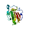
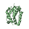
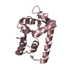
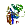
| Title | Cofilin homology domain of a yeast actin-binding protein ABP1P | ||||||
 Components Components | ACTIN-BINDING PROTEIN | ||||||
 Keywords Keywords | STRUCTURAL PROTEIN / cofilin homology domain / actin binding / New York SGX Research Center for Structural Genomics / NYSGXRC / Structural Genomics / PSI / Protein Structure Initiative | ||||||
| Function / homology |  Function and homology information Function and homology informationprotein localization to actin cortical patch / positive regulation of Arp2/3 complex-mediated actin nucleation / site of polarized growth / actin cortical patch assembly / actin cortical patch / regulation of actin filament polymerization / barbed-end actin filament capping / mating projection tip / cortical actin cytoskeleton / actin filament binding ...protein localization to actin cortical patch / positive regulation of Arp2/3 complex-mediated actin nucleation / site of polarized growth / actin cortical patch assembly / actin cortical patch / regulation of actin filament polymerization / barbed-end actin filament capping / mating projection tip / cortical actin cytoskeleton / actin filament binding / cell cortex / cytoplasm Similarity search - Function | ||||||
| Biological species |  | ||||||
| Method |  X-RAY DIFFRACTION / SYNCHROTRON / SIR, PHASED TRANSLATION FUNCTION, MOLECULAR REPLACEMENT / Resolution: 2.1 Å X-RAY DIFFRACTION / SYNCHROTRON / SIR, PHASED TRANSLATION FUNCTION, MOLECULAR REPLACEMENT / Resolution: 2.1 Å | ||||||
 Authors Authors | Strokopytov, B.V. / Fedorov, A.A. / Mahoney, N. / Drubin, D.G. / Almo, S.C. / Burley, S.K. / New York SGX Research Center for Structural Genomics (NYSGXRC) | ||||||
 Citation Citation | Journal: Acta Crystallogr.,Sect.D / Year: 2005 Title: Phased translation function revisited: structure solution of the cofilin-homology domain from yeast actin-binding protein 1 using six-dimensional searches. Authors: Strokopytov, B.V. / Fedorov, A. / Mahoney, N.M. / Kessels, M. / Drubin, D.G. / Almo, S.C. #1: Journal: Nat.Struct.Biol. / Year: 1997Title: Structure determination of yeast cofilin Authors: Fedorov, A.A. / Lappalainen, P. / Fedorov, E.V. / Drubin, D.G. / Almo, S.C. #2: Journal: Nature / Year: 1990Title: Homology of a yeast actin-binding protein to signal transduction proteins and myosin-1 Authors: Drubin, D.G. / Mulholland, J. / Zhu, Z. / Botstein, D. #3: Journal: J.Cell Biol. / Year: 1993Title: Cofilin is an essential component of the yeast cortical cytoskeleton Authors: Moon, A.L. / Janmey, P.A. / Louie, K.A. / Drubin, D.G. | ||||||
| History |
|
- Structure visualization
| Structure viewer | Molecule: MolmilJmol/JSmol |
|---|
- Downloads & links
-Download
| PDBx/mmCIF format | 1hqz.cif.gz | 252.7 KB | Display | PDBx/mmCIF format |
|---|---|---|---|---|
| PDB format | pdb1hqz.ent.gz | 205.8 KB | Display | PDB format |
| PDBx/mmJSON format | 1hqz.json.gz | Tree view | PDBx/mmJSON format | |
| Others |  Other downloads Other downloads |
-Validation report
| Arichive directory | https://data.pdbj.org/pub/pdb/validation_reports/hq/1hqzftp://data.pdbj.org/pub/pdb/validation_reports/hq/1hqz | HTTPS FTP |
|---|
-Related structure data
| Related structure data |  1cofS S: Starting model for refinement |
|---|---|
| Similar structure data | |
| Other databases |


-Links
 PDBj
PDBj
- Assembly
| Deposited unit | 
| ||||||||||
|---|---|---|---|---|---|---|---|---|---|---|---|
| 1 | 
| ||||||||||
| 2 | 
| ||||||||||
| 3 | 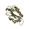
| ||||||||||
| 4 | 
| ||||||||||
| 5 | 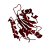
| ||||||||||
| 6 | 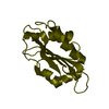
| ||||||||||
| 7 | 
| ||||||||||
| 8 | 
| ||||||||||
| 9 | 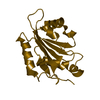
| ||||||||||
| 10 | 
| ||||||||||
| Unit cell |
| ||||||||||
| Components on special symmetry positions |
| ||||||||||
| Details | The biological assembly is a monomer |
-Components
| #1: Protein | Mass: 15638.401 Da / Num. of mol.: 9 / Fragment: N-TERMINAL DOMAIN, RESIDUES 1-141 Source method: isolated from a genetically manipulated source Source: (gene. exp.) Production host:  #2: Water | ChemComp-HOH / |  Mass: 18.015 Da / Num. of mol.: 516 / Source method: isolated from a natural source / Formula: H2O Mass: 18.015 Da / Num. of mol.: 516 / Source method: isolated from a natural source / Formula: H2O |
|---|
-Experimental details
-Experiment
| Experiment | Method: X-RAY DIFFRACTION / Number of used crystals: 1 |
|---|
- Sample preparation
| Crystal | Density Matthews: 2.22 Å3/Da / Density % sol: 43 % | ||||||||||||||||||
|---|---|---|---|---|---|---|---|---|---|---|---|---|---|---|---|---|---|---|---|
| Crystal grow | Temperature: 290 K / Method: vapor diffusion, hanging drop / pH: 5.6 Details: PEG 4000, sodium citrate, pH 5.6, VAPOR DIFFUSION, HANGING DROP at 290K | ||||||||||||||||||
| Crystal grow | *PLUS | ||||||||||||||||||
| Components of the solutions | *PLUS
|
-Data collection
| Diffraction | Mean temperature: 140 K |
|---|---|
| Diffraction source | Source: SYNCHROTRON / Site: NSLS  / Beamline: X9B / Wavelength: 1.04019 Å / Beamline: X9B / Wavelength: 1.04019 Å |
| Detector | Type: MARRESEARCH / Detector: IMAGE PLATE / Date: Nov 27, 1997 |
| Radiation | Protocol: SINGLE WAVELENGTH / Monochromatic (M) / Laue (L): M / Scattering type: x-ray |
| Radiation wavelength | Wavelength: 1.04019 Å / Relative weight: 1 |
| Reflection | Resolution: 2.1→15 Å / Num. obs: 68718 / % possible obs: 96.1 % / Observed criterion σ(F): 0 / Observed criterion σ(I): 0 / Redundancy: 2.1 % / Biso Wilson estimate: 29.5 Å2 / Rmerge(I) obs: 0.069 / Net I/σ(I): 9.9 |
| Reflection shell | Resolution: 2.1→2.23 Å / Redundancy: 1.4 % / Rmerge(I) obs: 0.397 / Num. unique all: 9605 / % possible all: 89.6 |
| Reflection | *PLUS Num. obs: 68796 / % possible obs: 96.6 % / Redundancy: 2.3 % |
| Reflection shell | *PLUS % possible obs: 92.6 % / Redundancy: 2.2 % / Rmerge(I) obs: 0.496 / Mean I/σ(I) obs: 1.9 |
- Processing
| Software |
| ||||||||||||||||||||||||||||||||||||||||
|---|---|---|---|---|---|---|---|---|---|---|---|---|---|---|---|---|---|---|---|---|---|---|---|---|---|---|---|---|---|---|---|---|---|---|---|---|---|---|---|---|---|
| Refinement | Method to determine structure: SIR, PHASED TRANSLATION FUNCTION, MOLECULAR REPLACEMENT Starting model: PDB ENTRY 1COF Resolution: 2.1→15 Å / Rfactor Rfree error: 0.003 / Isotropic thermal model: RESTRAINED / Cross valid method: THROUGHOUT / σ(F): 2 / Stereochemistry target values: Engh & Huber
| ||||||||||||||||||||||||||||||||||||||||
| Solvent computation | Solvent model: FLAT / Bsol: 50.2239 Å2 / ksol: 0.3641 e/Å3 | ||||||||||||||||||||||||||||||||||||||||
| Refine analyze |
| ||||||||||||||||||||||||||||||||||||||||
| Refinement step | Cycle: LAST / Resolution: 2.1→15 Å
| ||||||||||||||||||||||||||||||||||||||||
| Refine LS restraints |
| ||||||||||||||||||||||||||||||||||||||||
| LS refinement shell | Resolution: 2.1→2.23 Å / Rfactor Rfree error: 0.011 / Total num. of bins used: 6
| ||||||||||||||||||||||||||||||||||||||||
| Software | *PLUS Name: CNS / Version: 1 / Classification: refinement | ||||||||||||||||||||||||||||||||||||||||
| Refinement | *PLUS Lowest resolution: 20 Å / σ(F): 2 / % reflection Rfree: 10.1 % / Rfactor obs: 0.213 | ||||||||||||||||||||||||||||||||||||||||
| Solvent computation | *PLUS | ||||||||||||||||||||||||||||||||||||||||
| Displacement parameters | *PLUS | ||||||||||||||||||||||||||||||||||||||||
| Refine LS restraints | *PLUS
| ||||||||||||||||||||||||||||||||||||||||
| LS refinement shell | *PLUS Rfactor Rfree: 0.358 / % reflection Rfree: 10 % / Rfactor Rwork: 0.309 |
