 Movie
Movie Controller

[English] 日本語
 Yorodumi
- PDB-2pol: THREE-DIMENSIONAL STRUCTURE OF THE BETA SUBUNIT OF ESCHERICHIA CO... -
+ Open data

- Basic information
| Entry | Database: PDB / ID: 2pol | ||||||
|---|---|---|---|---|---|---|---|
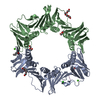
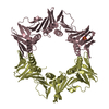
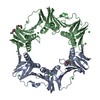
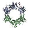
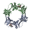
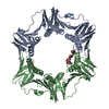
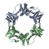
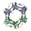
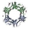
| Title | THREE-DIMENSIONAL STRUCTURE OF THE BETA SUBUNIT OF ESCHERICHIA COLI DNA POLYMERASE III HOLOENZYME: A SLIDING DNA CLAMP | ||||||
 Components Components | DNA POLYMERASE III (BETA SUBUNIT) | ||||||
 Keywords Keywords | NUCLEOTIDYLTRANSFERASE | ||||||
| Function / homology |  Function and homology information Function and homology informationreplication inhibiting complex / Hda-beta clamp complex / bacterial-type DNA replication / DNA polymerase III complex / replisome / regulation of DNA-templated DNA replication initiation / DNA strand elongation involved in DNA replication / error-prone translesion synthesis / 3'-5' exonuclease activity / negative regulation of DNA-templated DNA replication initiation ...replication inhibiting complex / Hda-beta clamp complex / bacterial-type DNA replication / DNA polymerase III complex / replisome / regulation of DNA-templated DNA replication initiation / DNA strand elongation involved in DNA replication / error-prone translesion synthesis / 3'-5' exonuclease activity / negative regulation of DNA-templated DNA replication initiation / DNA-templated DNA replication / DNA-directed DNA polymerase activity / DNA damage response / protein homodimerization activity / DNA binding / identical protein binding / cytosol Similarity search - Function | ||||||
| Biological species |  | ||||||
| Method |  X-RAY DIFFRACTION / Resolution: 2.5 Å X-RAY DIFFRACTION / Resolution: 2.5 Å | ||||||
 Authors Authors | Kong, X.-P. / Kuriyan, J. | ||||||
 Citation Citation | Journal: Cell(Cambridge,Mass.) / Year: 1992 Title: Three-dimensional structure of the beta subunit of E. coli DNA polymerase III holoenzyme: a sliding DNA clamp. Authors: Kong, X.P. / Onrust, R. / O'Donnell, M. / Kuriyan, J. | ||||||
| History |
|
- Structure visualization
| Structure viewer | Molecule: MolmilJmol/JSmol |
|---|
- Downloads & links
-Download
| PDBx/mmCIF format | 2pol.cif.gz | 151.8 KB | Display | PDBx/mmCIF format |
|---|---|---|---|---|
| PDB format | pdb2pol.ent.gz | 121.4 KB | Display | PDB format |
| PDBx/mmJSON format | 2pol.json.gz | Tree view | PDBx/mmJSON format | |
| Others |  Other downloads Other downloads |
-Validation report
| Arichive directory | https://data.pdbj.org/pub/pdb/validation_reports/po/2polftp://data.pdbj.org/pub/pdb/validation_reports/po/2pol | HTTPS FTP |
|---|
-Related structure data
| Similar structure data |
|---|


-Links
 PDBj
PDBj
- Assembly
| Deposited unit | 
| ||||||||
|---|---|---|---|---|---|---|---|---|---|
| 1 |
| ||||||||
| Unit cell |
|
-Components
| #1: Protein | Mass: 40630.508 Da / Num. of mol.: 2 Source method: isolated from a genetically manipulated source Source: (gene. exp.) #2: Water | ChemComp-HOH / |  Mass: 18.015 Da / Num. of mol.: 150 / Source method: isolated from a natural source / Formula: H2O Mass: 18.015 Da / Num. of mol.: 150 / Source method: isolated from a natural source / Formula: H2O |
|---|
-Experimental details
-Experiment
| Experiment | Method: X-RAY DIFFRACTION |
|---|
- Sample preparation
| Crystal | Density Matthews: 2.54 Å3/Da / Density % sol: 51.65 % | ||||||||||||||||||||||||
|---|---|---|---|---|---|---|---|---|---|---|---|---|---|---|---|---|---|---|---|---|---|---|---|---|---|
| Crystal grow | *PLUS Temperature: 21 ℃ / pH: 6 / Method: vapor diffusion, hanging drop | ||||||||||||||||||||||||
| Components of the solutions | *PLUS
|
-Data collection
| Reflection | *PLUS Highest resolution: 2.4 Å / Num. obs: 30957 / % possible obs: 94.4 % / Num. measured all: 101590 / Rmerge(I) obs: 0.0729 |
|---|
- Processing
| Software |
| ||||||||||||||||||||||||||||||||||||||||||||||||||||||||||||
|---|---|---|---|---|---|---|---|---|---|---|---|---|---|---|---|---|---|---|---|---|---|---|---|---|---|---|---|---|---|---|---|---|---|---|---|---|---|---|---|---|---|---|---|---|---|---|---|---|---|---|---|---|---|---|---|---|---|---|---|---|---|
| Refinement | Rfactor Rwork: 0.184 / Rfactor obs: 0.184 / Highest resolution: 2.5 Å | ||||||||||||||||||||||||||||||||||||||||||||||||||||||||||||
| Refinement step | Cycle: LAST / Highest resolution: 2.5 Å
| ||||||||||||||||||||||||||||||||||||||||||||||||||||||||||||
| Refine LS restraints |
| ||||||||||||||||||||||||||||||||||||||||||||||||||||||||||||
| Software | *PLUS Name: X-PLOR / Classification: refinement | ||||||||||||||||||||||||||||||||||||||||||||||||||||||||||||
| Refinement | *PLUS Highest resolution: 2.5 Å / Num. reflection obs: 27614 / σ(F): 2 / Rfactor obs: 0.184 | ||||||||||||||||||||||||||||||||||||||||||||||||||||||||||||
| Solvent computation | *PLUS | ||||||||||||||||||||||||||||||||||||||||||||||||||||||||||||
| Displacement parameters | *PLUS | ||||||||||||||||||||||||||||||||||||||||||||||||||||||||||||
| Refine LS restraints | *PLUS Type: x_angle_d / Dev ideal: 3.45 |
