replication inhibiting complex / Hda-beta clamp complex / bacterial-type DNA replication / DNA polymerase III complex / replisome / regulation of DNA-templated DNA replication initiation / DNA strand elongation involved in DNA replication / error-prone translesion synthesis / 3'-5' exonuclease activity / negative regulation of DNA-templated DNA replication initiation ...replication inhibiting complex / Hda-beta clamp complex / bacterial-type DNA replication / DNA polymerase III complex / replisome / regulation of DNA-templated DNA replication initiation / DNA strand elongation involved in DNA replication / error-prone translesion synthesis / 3'-5' exonuclease activity / negative regulation of DNA-templated DNA replication initiation / DNA-templated DNA replication / DNA-directed DNA polymerase activity / DNA damage response / protein homodimerization activity / DNA binding / identical protein binding / cytosol Similarity search - Function
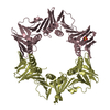
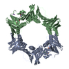
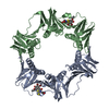
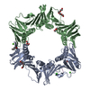
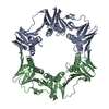
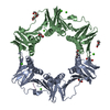
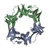
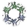
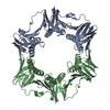
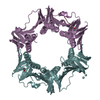
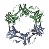
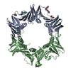
DNA Polymerase III; Chain A, domain 2 / DNA Polymerase III, subunit A, domain 2 / DNA polymerase III, beta sliding clamp / DNA polymerase III, beta sliding clamp, N-terminal / DNA polymerase III, beta sliding clamp, C-terminal / DNA polymerase III, beta sliding clamp, central / DNA polymerase III beta subunit, N-terminal domain / DNA polymerase III beta subunit, central domain / DNA polymerase III beta subunit, C-terminal domain / DNA polymerase III beta subunit ...DNA Polymerase III; Chain A, domain 2 / DNA Polymerase III, subunit A, domain 2 / DNA polymerase III, beta sliding clamp / DNA polymerase III, beta sliding clamp, N-terminal / DNA polymerase III, beta sliding clamp, C-terminal / DNA polymerase III, beta sliding clamp, central / DNA polymerase III beta subunit, N-terminal domain / DNA polymerase III beta subunit, central domain / DNA polymerase III beta subunit, C-terminal domain / DNA polymerase III beta subunit / : / Roll / Alpha Beta Similarity search - Domain/homology
 Movie
Movie Controller


 Open data
 Basic information
 Components
 Keywords
 Function and homology information
 X-RAY DIFFRACTION /
  Authors
 Citation
 Structure visualization
 Downloads & links
 Other downloads
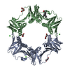


 PDBj
PDBj
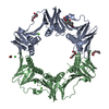
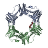
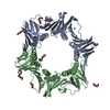
 Assembly


 Mass: 150.173 Da / Num. of mol.: 2 / Source method: obtained synthetically / Formula: C6H14O4
 Mass: 106.120 Da / Num. of mol.: 4 / Source method: obtained synthetically / Formula: C4H10O3
 Mass: 35.453 Da / Num. of mol.: 3 / Source method: obtained synthetically / Formula: Cl
 Mass: 273.714 Da / Num. of mol.: 1 / Source method: obtained synthetically / Formula: C15H12ClNO2
 Mass: 40.078 Da / Num. of mol.: 1 / Source method: obtained synthetically / Formula: Ca
 Sample preparation
 / Beamline: MX1 / Wavelength: 0.95 Å
 Processing