 Movie
Movie Controller

[English] 日本語
 Yorodumi
- PDB-1uin: Crystal Structure of Threonine Synthase from Thermus Thermophilus... -
+ Open data

- Basic information
| Entry | Database: PDB / ID: 1uin | ||||||
|---|---|---|---|---|---|---|---|
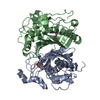
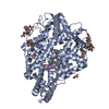
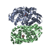
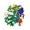
| Title | Crystal Structure of Threonine Synthase from Thermus Thermophilus HB8, Trigonal Crystal Form | ||||||
 Components Components | Threonine Synthase | ||||||
 Keywords Keywords | LYASE / PLP-dependent enzyme / RIKEN Structural Genomics/Proteomics Initiative / RSGI / Structural Genomics | ||||||
| Function / homology |  Function and homology information Function and homology informationthreonine synthase / threonine synthase activity / threonine deaminase activity / L-serine ammonia-lyase activity / L-threonine catabolic process / L-serine catabolic process / L-threonine biosynthetic process / : / pyridoxal phosphate binding Similarity search - Function | ||||||
| Biological species |   Thermus thermophilus (bacteria) Thermus thermophilus (bacteria) | ||||||
| Method |  X-RAY DIFFRACTION / SYNCHROTRON / MIR / Resolution: 2.25 Å X-RAY DIFFRACTION / SYNCHROTRON / MIR / Resolution: 2.25 Å | ||||||
 Authors Authors | Omi, R. / RIKEN Structural Genomics/Proteomics Initiative (RSGI) | ||||||
 Citation Citation | Journal: J.BIOL.CHEM. / Year: 2003 Title: Crystal Structures of Threonine Synthase from Thermus thermophilus HB8: Conformational change, substrate recognition, and mechanism. Authors: Omi, R. / Masaru, G. / Miyahara, I. / Mizuguchi, H. / Hayashi, H. / Kagamiyama, H. / Hirotsu, K. | ||||||
| History |
|
- Structure visualization
| Structure viewer | Molecule: MolmilJmol/JSmol |
|---|
- Downloads & links
-Download
| PDBx/mmCIF format | 1uin.cif.gz | 139.1 KB | Display | PDBx/mmCIF format |
|---|---|---|---|---|
| PDB format | pdb1uin.ent.gz | 110.3 KB | Display | PDB format |
| PDBx/mmJSON format | 1uin.json.gz | Tree view | PDBx/mmJSON format | |
| Others |  Other downloads Other downloads |
-Validation report
| Arichive directory | https://data.pdbj.org/pub/pdb/validation_reports/ui/1uinftp://data.pdbj.org/pub/pdb/validation_reports/ui/1uin | HTTPS FTP |
|---|
-Related structure data


-Links
 PDBj
PDBj
- Assembly
| Deposited unit | 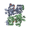
| ||||||||
|---|---|---|---|---|---|---|---|---|---|
| 1 |
| ||||||||
| 2 | 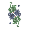
| ||||||||
| Unit cell |
|
-Components
| #1: Protein | Mass: 37054.656 Da / Num. of mol.: 2 Source method: isolated from a genetically manipulated source Source: (gene. exp.) Thermus thermophilus (bacteria) / Gene: HB8 / Plasmid: pET20b(+) / Production host: #2: Chemical |   Mass: 96.063 Da / Num. of mol.: 3 / Source method: obtained synthetically / Formula: SO4 Mass: 96.063 Da / Num. of mol.: 3 / Source method: obtained synthetically / Formula: SO4#3: Chemical |   Mass: 247.142 Da / Num. of mol.: 2 / Source method: obtained synthetically / Formula: C8H10NO6P Mass: 247.142 Da / Num. of mol.: 2 / Source method: obtained synthetically / Formula: C8H10NO6P#4: Water | ChemComp-HOH / |  Mass: 18.015 Da / Num. of mol.: 211 / Source method: isolated from a natural source / Formula: H2O Mass: 18.015 Da / Num. of mol.: 211 / Source method: isolated from a natural source / Formula: H2O |
|---|
-Experimental details
-Experiment
| Experiment | Method: X-RAY DIFFRACTION / Number of used crystals: 1 |
|---|
- Sample preparation
| Crystal | Density Matthews: 3.71 Å3/Da / Density % sol: 66.63 % | ||||||||||||||||||
|---|---|---|---|---|---|---|---|---|---|---|---|---|---|---|---|---|---|---|---|
| Crystal grow | Temperature: 293 K / Method: vapor diffusion, hanging drop / pH: 6.5 Details: Ammonium Sulfate, Mes, pH 6.5, VAPOR DIFFUSION, HANGING DROP, temperature 293K | ||||||||||||||||||
| Crystal grow | *PLUS Method: vapor diffusion, hanging drop | ||||||||||||||||||
| Components of the solutions | *PLUS
|
-Data collection
| Diffraction | Mean temperature: 100 K |
|---|---|
| Diffraction source | Source: SYNCHROTRON / Site: SPring-8  / Beamline: BL40B2 / Wavelength: 1 Å / Beamline: BL40B2 / Wavelength: 1 Å |
| Detector | Type: ADSC QUANTUM 4 / Detector: CCD / Date: Jan 28, 2003 |
| Radiation | Protocol: SINGLE WAVELENGTH / Monochromatic (M) / Laue (L): M / Scattering type: x-ray |
| Radiation wavelength | Wavelength: 1 Å / Relative weight: 1 |
| Reflection | Resolution: 2.25→20 Å / Num. all: 53148 / Num. obs: 53148 / % possible obs: 99.7 % |
| Reflection shell | Resolution: 2.25→2.33 Å / % possible all: 99.4 |
| Reflection | *PLUS Num. measured all: 486914 / Rmerge(I) obs: 0.08 |
| Reflection shell | *PLUS % possible obs: 99.4 % / Rmerge(I) obs: 0.323 |
- Processing
| Software |
| ||||||||||||||||||||
|---|---|---|---|---|---|---|---|---|---|---|---|---|---|---|---|---|---|---|---|---|---|
| Refinement | Method to determine structure: MIR / Resolution: 2.25→10 Å / σ(F): 2
| ||||||||||||||||||||
| Refine analyze | Luzzati coordinate error obs: 0.24 Å / Luzzati d res low obs: 5 Å / Luzzati sigma a obs: 0.16 Å | ||||||||||||||||||||
| Refinement step | Cycle: LAST / Resolution: 2.25→10 Å
| ||||||||||||||||||||
| Refine LS restraints |
| ||||||||||||||||||||
| Refinement | *PLUS Rfactor Rwork: 0.201 | ||||||||||||||||||||
| Solvent computation | *PLUS | ||||||||||||||||||||
| Displacement parameters | *PLUS | ||||||||||||||||||||
| Refine LS restraints | *PLUS
| ||||||||||||||||||||
| LS refinement shell | *PLUS Rfactor Rfree: 0.226 / Rfactor Rwork: 0.211 |
