 ムービー
ムービー コントローラー


+ データを開く

- 基本情報
基本情報
| 登録情報 | データベース: PDB / ID: 1ugh | ||||||
|---|---|---|---|---|---|---|---|
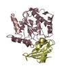
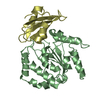
| タイトル | CRYSTAL STRUCTURE OF HUMAN URACIL-DNA GLYCOSYLASE IN COMPLEX WITH A PROTEIN INHIBITOR: PROTEIN MIMICRY OF DNA | ||||||
 要素 要素 |
| ||||||
 キーワード キーワード | GLYCOSYLASE / ENZYME-INHIBITOR COMPLEX | ||||||
| 機能・相同性 |  機能・相同性情報 機能・相同性情報base-excision repair, AP site formation via deaminated base removal / uracil-DNA glycosylase / depyrimidination / Displacement of DNA glycosylase by APEX1 / single strand break repair / isotype switching / uracil DNA N-glycosylase activity / ribosomal small subunit binding / somatic hypermutation of immunoglobulin genes / Recognition and association of DNA glycosylase with site containing an affected pyrimidine ...base-excision repair, AP site formation via deaminated base removal / uracil-DNA glycosylase / depyrimidination / Displacement of DNA glycosylase by APEX1 / single strand break repair / isotype switching / uracil DNA N-glycosylase activity / ribosomal small subunit binding / somatic hypermutation of immunoglobulin genes / Recognition and association of DNA glycosylase with site containing an affected pyrimidine / Cleavage of the damaged pyrimidine / Chromatin modifications during the maternal to zygotic transition (MZT) / base-excision repair / damaged DNA binding / negative regulation of apoptotic process / mitochondrion / nucleoplasm / nucleus 類似検索 - 分子機能 | ||||||
| 生物種 |  Homo sapiens (ヒト) Homo sapiens (ヒト) Bacillus phage PBS2 (ファージ) Bacillus phage PBS2 (ファージ) | ||||||
| 手法 |  X線回折 / シンクロトロン / 分子置換 / 解像度: 1.9 Å X線回折 / シンクロトロン / 分子置換 / 解像度: 1.9 Å | ||||||
 データ登録者 データ登録者 | Mol, C.D. / Arvai, A.S. / Sanderson, R.J. / Slupphaug, G. / Kavli, B. / Krokan, H.E. / Mosbaugh, D.W. / Tainer, J.A. | ||||||
 引用 引用 | ジャーナル: Cell(Cambridge,Mass.) / 年: 1995 タイトル: Crystal structure of human uracil-DNA glycosylase in complex with a protein inhibitor: protein mimicry of DNA. 著者: Mol, C.D. / Arvai, A.S. / Sanderson, R.J. / Slupphaug, G. / Kavli, B. / Krokan, H.E. / Mosbaugh, D.W. / Tainer, J.A. #1: ジャーナル: J.Mol.Biol. / 年: 1999タイトル: Protein mimicry of DNA from crystal structures of the uracil-DNA glycosylase inhibitor protein and its complex with Escherichia coli uracil-DNA glycosylase. 著者: Putnam, C.D. / Shroyer, M.J. / Lundquist, A.J. / Mol, C.D. / Arvai, A.S. / Mosbaugh, D.W. / Tainer, J.A. #2: ジャーナル: Embo J. / 年: 1998タイトル: Base excision repair initiation revealed by crystal structures and binding kinetics of human uracil-DNA glycosylase with DNA. 著者: Parikh, S.S. / Mol, C.D. / Slupphaug, G. / Bharati, S. / Krokan, H.E. / Tainer, J.A. #3: ジャーナル: Nature / 年: 1996タイトル: A nucleotide-flipping mechanism from the structure of human uracil-DNA glycosylase bound to DNA. 著者: Slupphaug, G. / Mol, C.D. / Kavli, B. / Arvai, A.S. / Krokan, H.E. / Tainer, J.A. #4: ジャーナル: Cell(Cambridge,Mass.) / 年: 1995 タイトル: Crystal structure and mutational analysis of human uracil-DNA glycosylase: structural basis for specificity and catalysis. 著者: Mol, C.D. / Arvai, A.S. / Slupphaug, G. / Kavli, B. / Alseth, I. / Krokan, H.E. / Tainer, J.A. | ||||||
| 履歴 |
|
- 構造の表示
構造の表示
| 構造ビューア | 分子: MolmilJmol/JSmol |
|---|
- ダウンロードとリンク
-ダウンロード
| PDBx/mmCIF形式 | 1ugh.cif.gz | 78.5 KB | 表示 | PDBx/mmCIF形式 |
|---|---|---|---|---|
| PDB形式 | pdb1ugh.ent.gz | 57.7 KB | 表示 | PDB形式 |
| PDBx/mmJSON形式 | 1ugh.json.gz | ツリー表示 | PDBx/mmJSON形式 | |
| その他 |  その他のダウンロード その他のダウンロード |
-検証レポート
| 文書・要旨 | 1ugh_validation.pdf.gz | 431.9 KB | 表示 | wwPDB検証レポート |
|---|---|---|---|---|
| 文書・詳細版 | 1ugh_full_validation.pdf.gz | 435.8 KB | 表示 | |
| XML形式データ | 1ugh_validation.xml.gz | 15.5 KB | 表示 | |
| CIF形式データ | 1ugh_validation.cif.gz | 21.6 KB | 表示 | |
| アーカイブディレクトリ | https://data.pdbj.org/pub/pdb/validation_reports/ug/1ughftp://data.pdbj.org/pub/pdb/validation_reports/ug/1ugh | HTTPS FTP |
-関連構造データ
| 関連構造データ | 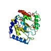 1akzS S: 精密化の開始モデル |
|---|---|
| 類似構造データ |


-リンク
 PDBj
PDBj
- 集合体
集合体
| 登録構造単位 | 
| ||||||||||
|---|---|---|---|---|---|---|---|---|---|---|---|
| 1 |
| ||||||||||
| 単位格子 |
|
-要素
| #1: タンパク質 | 分子量: 25544.137 Da / 分子数: 1 / 変異: P82M, V83E, G84F / 由来タイプ: 組換発現 / 由来: (組換発現) Homo sapiens (ヒト) / 発現宿主:  |
|---|---|
| #2: タンパク質 | 分子量: 9250.373 Da / 分子数: 1 / 由来タイプ: 組換発現 / 由来: (組換発現) Bacillus phage PBS2 (ファージ) / 発現宿主: |
| #3: 水 | ChemComp-HOH /  分子量: 18.015 Da / 分子数: 185 / 由来タイプ: 天然 / 式: H2O 分子量: 18.015 Da / 分子数: 185 / 由来タイプ: 天然 / 式: H2O |
-実験情報
-実験
| 実験 | 手法: X線回折 / 使用した結晶の数: 1 |
|---|
- 試料調製
試料調製
| 結晶 | マシュー密度: 2.3 Å3/Da / 溶媒含有率: 47 % | |||||||||||||||||||||||||
|---|---|---|---|---|---|---|---|---|---|---|---|---|---|---|---|---|---|---|---|---|---|---|---|---|---|---|
| 結晶化 | pH: 5 / 詳細: pH 5.0 | |||||||||||||||||||||||||
| 溶液の組成 |
| |||||||||||||||||||||||||
| 結晶化 | *PLUS 手法: 蒸気拡散法, ハンギングドロップ法詳細: drop consists of equal volume of protein and reservoir solutions | |||||||||||||||||||||||||
| 溶液の組成 | *PLUS
|
-データ収集
| 回折 | 平均測定温度: 277 K |
|---|---|
| 放射光源 | 由来: シンクロトロン / サイト: SSRL  / ビームライン: BL7-1 / 波長: 1.07 / ビームライン: BL7-1 / 波長: 1.07 |
| 検出器 | タイプ: MARRESEARCH / 検出器: IMAGE PLATE / 日付: 1995年3月15日 |
| 放射 | プロトコル: SINGLE WAVELENGTH / 単色(M)・ラウエ(L): M / 散乱光タイプ: x-ray |
| 放射波長 | 波長: 1.07 Å / 相対比: 1 |
| 反射 | 解像度: 1.9→8 Å / Num. obs: 21979 / % possible obs: 90 % / 冗長度: 1.92 % / Biso Wilson estimate: 21.4 Å2 / Rsym value: 0.047 / Net I/σ(I): 9.7 |
| 反射 シェル | 解像度: 1.9→1.97 Å / Rsym value: 0.224 / % possible all: 76 |
| 反射 | *PLUS Rmerge(I) obs: 0.047 |
| 反射 シェル | *PLUS % possible obs: 76 % / Rmerge(I) obs: 0.224 |
- 解析
解析
| ソフトウェア |
| ||||||||||||||||||||||||||||||||||||||||||||||||||||||||||||
|---|---|---|---|---|---|---|---|---|---|---|---|---|---|---|---|---|---|---|---|---|---|---|---|---|---|---|---|---|---|---|---|---|---|---|---|---|---|---|---|---|---|---|---|---|---|---|---|---|---|---|---|---|---|---|---|---|---|---|---|---|---|
| 精密化 | 構造決定の手法: 分子置換 開始モデル: PDB ENTRY 1AKZ 解像度: 1.9→8 Å / 交差検証法: THROUGHOUT / σ(F): 3 詳細: UGI RESIDUES MET1 AND THR2 ARE DISORDERED AND NOT SEEN IN THE ELECTRON DENSITY
| ||||||||||||||||||||||||||||||||||||||||||||||||||||||||||||
| 原子変位パラメータ | Biso mean: 35 Å2
| ||||||||||||||||||||||||||||||||||||||||||||||||||||||||||||
| 精密化ステップ | サイクル: LAST / 解像度: 1.9→8 Å
| ||||||||||||||||||||||||||||||||||||||||||||||||||||||||||||
| 拘束条件 |
| ||||||||||||||||||||||||||||||||||||||||||||||||||||||||||||
| LS精密化 シェル | 解像度: 1.9→1.99 Å / Total num. of bins used: 8
| ||||||||||||||||||||||||||||||||||||||||||||||||||||||||||||
| Xplor file |
| ||||||||||||||||||||||||||||||||||||||||||||||||||||||||||||
| ソフトウェア | *PLUS 名称: X-PLOR / バージョン: 3.851 / 分類: refinement | ||||||||||||||||||||||||||||||||||||||||||||||||||||||||||||
| 精密化 | *PLUS Rfactor obs: 0.198 | ||||||||||||||||||||||||||||||||||||||||||||||||||||||||||||
| 溶媒の処理 | *PLUS | ||||||||||||||||||||||||||||||||||||||||||||||||||||||||||||
| 原子変位パラメータ | *PLUS | ||||||||||||||||||||||||||||||||||||||||||||||||||||||||||||
| 拘束条件 | *PLUS
| ||||||||||||||||||||||||||||||||||||||||||||||||||||||||||||
| LS精密化 シェル | *PLUS Rfactor obs: 0.3394 |
