 Movie
Movie Controller

[English] 日本語
 Yorodumi
- PDB-1uex: Crystal structure of von Willebrand Factor A1 domain complexed wi... -
+ Open data

- Basic information
| Entry | Database: PDB / ID: 1uex | ||||||
|---|---|---|---|---|---|---|---|
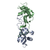
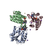
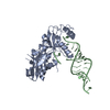
| Title | Crystal structure of von Willebrand Factor A1 domain complexed with snake venom bitiscetin | ||||||
 Components Components |
| ||||||
 Keywords Keywords | TOXIN/BLOOD CLOTTING / C-type lectin heterodimer / TOXIN-BLOOD CLOTTING COMPLEX | ||||||
| Function / homology |  Function and homology information Function and homology informationDefective VWF binding to collagen type I / Enhanced cleavage of VWF variant by ADAMTS13 / Defective VWF cleavage by ADAMTS13 variant / Defective F8 binding to von Willebrand factor / Enhanced binding of GP1BA variant to VWF multimer:collagen / Defective binding of VWF variant to GPIb:IX:V / Weibel-Palade body / hemostasis / platelet alpha granule / Platelet Adhesion to exposed collagen ...Defective VWF binding to collagen type I / Enhanced cleavage of VWF variant by ADAMTS13 / Defective VWF cleavage by ADAMTS13 variant / Defective F8 binding to von Willebrand factor / Enhanced binding of GP1BA variant to VWF multimer:collagen / Defective binding of VWF variant to GPIb:IX:V / Weibel-Palade body / hemostasis / platelet alpha granule / Platelet Adhesion to exposed collagen / extracellular matrix structural constituent / GP1b-IX-V activation signalling / p130Cas linkage to MAPK signaling for integrins / Defective F8 cleavage by thrombin / Platelet Aggregation (Plug Formation) / cell-substrate adhesion / GRB2:SOS provides linkage to MAPK signaling for Integrins / positive regulation of intracellular signal transduction / immunoglobulin binding / Integrin cell surface interactions / collagen binding / : / Integrin signaling / platelet alpha granule lumen / Signaling by high-kinase activity BRAF mutants / platelet activation / MAP2K and MAPK activation / response to wounding / integrin binding / blood coagulation / Signaling by RAF1 mutants / Signaling by moderate kinase activity BRAF mutants / Paradoxical activation of RAF signaling by kinase inactive BRAF / Signaling downstream of RAS mutants / Signaling by BRAF and RAF1 fusions / Platelet degranulation / toxin activity / protein-folding chaperone binding / extracellular matrix / protease binding / cell adhesion / endoplasmic reticulum / : / extracellular exosome / extracellular region / identical protein binding Similarity search - Function | ||||||
| Biological species |  Homo sapiens (human) Homo sapiens (human) Bitis arietans (puff adder) Bitis arietans (puff adder) | ||||||
| Method |  X-RAY DIFFRACTION / SYNCHROTRON / MOLECULAR REPLACEMENT / Resolution: 2.85 Å X-RAY DIFFRACTION / SYNCHROTRON / MOLECULAR REPLACEMENT / Resolution: 2.85 Å | ||||||
 Authors Authors | Maita, N. / Nishio, K. / Nishimoto, E. / Matsui, T. / Shikamoto, Y. / Morita, T. / Sadler, J.E. / Mizuno, H. | ||||||
 Citation Citation | Journal: J.Biol.Chem. / Year: 2003 Title: Crystal structure of von Willebrand factor A1 domain complexed with snake venom, bitiscetin. Insight into glycoprotein Ibalpha binding mechanism induced by snake venom proteins. Authors: Maita, N. / Nishio, K. / Nishimoto, E. / Matsui, T. / Shikamoto, Y. / Morita, T. / Sadler, J.E. / Mizuno, H. | ||||||
| History |
|
- Structure visualization
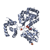
| Structure viewer | Molecule: MolmilJmol/JSmol |
|---|
- Downloads & links
-Download
| PDBx/mmCIF format | 1uex.cif.gz | 103.4 KB | Display | PDBx/mmCIF format |
|---|---|---|---|---|
| PDB format | pdb1uex.ent.gz | 79.7 KB | Display | PDB format |
| PDBx/mmJSON format | 1uex.json.gz | Tree view | PDBx/mmJSON format | |
| Others |  Other downloads Other downloads |
-Validation report
| Arichive directory | https://data.pdbj.org/pub/pdb/validation_reports/ue/1uexftp://data.pdbj.org/pub/pdb/validation_reports/ue/1uex | HTTPS FTP |
|---|
-Related structure data


-Links
 PDBj
PDBj


- Assembly
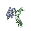
| Deposited unit | 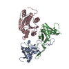
| ||||||||
|---|---|---|---|---|---|---|---|---|---|
| 1 |
| ||||||||
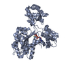
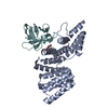
| Unit cell |
|
-Components
| #1: Protein | Mass: 14953.644 Da / Num. of mol.: 1 / Source method: isolated from a natural source / Source: (natural) Bitis arietans (puff adder) / Secretion: venom / References: GenBank: 2134244, UniProt: Q7LZK5*PLUS |
|---|---|
| #2: Protein | Mass: 14822.174 Da / Num. of mol.: 1 / Source method: isolated from a natural source / Source: (natural) Bitis arietans (puff adder) / Secretion: venom / References: GenBank: 2134245, UniProt: Q7LZK8*PLUS |
| #3: Protein | Mass: 23889.627 Da / Num. of mol.: 1 / Fragment: A1 domain Source method: isolated from a genetically manipulated source Source: (gene. exp.) Homo sapiens (human) / Plasmid: pET11b / Species (production host): Escherichia coli / Production host:  |
| #4: Water | ChemComp-HOH /  Mass: 18.015 Da / Num. of mol.: 40 / Source method: isolated from a natural source / Formula: H2O Mass: 18.015 Da / Num. of mol.: 40 / Source method: isolated from a natural source / Formula: H2O |
| Has protein modification | Y |
-Experimental details
-Experiment
| Experiment | Method: X-RAY DIFFRACTION / Number of used crystals: 1 |
|---|
- Sample preparation
| Crystal | Density Matthews: 2.06 Å3/Da / Density % sol: 39.8 % | ||||||||||||||||||||||||||||||||||||||||||
|---|---|---|---|---|---|---|---|---|---|---|---|---|---|---|---|---|---|---|---|---|---|---|---|---|---|---|---|---|---|---|---|---|---|---|---|---|---|---|---|---|---|---|---|
| Crystal grow | Temperature: 283 K / Method: vapor diffusion, sitting drop / pH: 7.8 Details: 10% PEG 6000, 2% PEG-MME 550, 5% MPD, pH 7.8, VAPOR DIFFUSION, SITTING DROP, temperature 283K | ||||||||||||||||||||||||||||||||||||||||||
| Crystal grow | *PLUS pH: 7.5 / Method: vapor diffusion, sitting drop | ||||||||||||||||||||||||||||||||||||||||||
| Components of the solutions | *PLUS
|
-Data collection
| Diffraction | Mean temperature: 100 K |
|---|---|
| Diffraction source | Source: SYNCHROTRON / Site: Photon Factory  / Beamline: BL-6B / Wavelength: 1 Å / Beamline: BL-6B / Wavelength: 1 Å |
| Detector | Type: RIGAKU RAXIS IV / Detector: IMAGE PLATE / Date: Oct 13, 2002 |
| Radiation | Protocol: SINGLE WAVELENGTH / Monochromatic (M) / Laue (L): M / Scattering type: x-ray |
| Radiation wavelength | Wavelength: 1 Å / Relative weight: 1 |
| Reflection | Resolution: 2.85→30 Å / Num. obs: 9672 / % possible obs: 91.6 % / Redundancy: 5.6 % / Rsym value: 0.107 |
| Reflection shell | Resolution: 2.85→2.99 Å / Redundancy: 5.7 % / Mean I/σ(I) obs: 2.3 / Rsym value: 0.31 / % possible all: 91.6 |
| Reflection | *PLUS Num. obs: 9055 / Num. measured all: 51631 / Rmerge(I) obs: 0.107 |
| Reflection shell | *PLUS % possible obs: 91.6 % / Num. unique obs: 1456 / Num. measured obs: 8285 / Rmerge(I) obs: 0.311 |
- Processing
| Software |
| ||||||||||||||||||||
|---|---|---|---|---|---|---|---|---|---|---|---|---|---|---|---|---|---|---|---|---|---|
| Refinement | Method to determine structure: MOLECULAR REPLACEMENT Starting model: PDB ENTRY 1JWI and 1AUQ Resolution: 2.85→20 Å / σ(F): 0 / Stereochemistry target values: Engh & Huber
| ||||||||||||||||||||
| Refinement step | Cycle: LAST / Resolution: 2.85→20 Å
| ||||||||||||||||||||
| Refine LS restraints |
| ||||||||||||||||||||
| Refinement | *PLUS % reflection Rfree: 12 % | ||||||||||||||||||||
| Solvent computation | *PLUS | ||||||||||||||||||||
| Displacement parameters | *PLUS | ||||||||||||||||||||
| Refine LS restraints | *PLUS
|
