 Movie
Movie Controller


+ Open data

- Basic information
| Entry | Database: PDB / ID: 1ue3 | ||||||||||||||||||
|---|---|---|---|---|---|---|---|---|---|---|---|---|---|---|---|---|---|---|---|
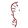
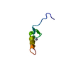
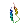
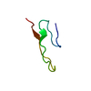
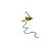
| Title | Crystal structure of d(GCGAAAGC) containing hexaamminecobalt | ||||||||||||||||||
 Components Components | 5'-D(* Keywords KeywordsDNA / base-intercalated duplex / mini-hairpin structure / sheared G:A pair / zipper-like duplex | Function / homology | COBALT HEXAMMINE(III) / DNA |  Function and homology information Function and homology informationMethod |  X-RAY DIFFRACTION / SYNCHROTRON / MOLECULAR REPLACEMENT / Resolution: 2.15 Å X-RAY DIFFRACTION / SYNCHROTRON / MOLECULAR REPLACEMENT / Resolution: 2.15 Å  Authors AuthorsSunami, T. / Kondo, J. / Hirao, I. / Watanabe, K. / Miura, K. / Takenaka, A. |  CitationJournal: Acta Crystallogr.,Sect.D / Year: 2004 CitationJournal: Acta Crystallogr.,Sect.D / Year: 2004Title: Structure of d(GCGAAAGC) (hexagonal form): a base-intercalated duplex as a stable structure. Authors: Sunami, T. / Kondo, J. / Hirao, I. / Watanabe, K. / Miura, K.I. / Takenaka, A. History |
|
- Structure visualization
| Structure viewer | Molecule: MolmilJmol/JSmol |
|---|
- Downloads & links
-Download
| PDBx/mmCIF format | 1ue3.cif.gz | 16.7 KB | Display | PDBx/mmCIF format |
|---|---|---|---|---|
| PDB format | pdb1ue3.ent.gz | 9.4 KB | Display | PDB format |
| PDBx/mmJSON format | 1ue3.json.gz | Tree view | PDBx/mmJSON format | |
| Others |  Other downloads Other downloads |
-Validation report
| Arichive directory | https://data.pdbj.org/pub/pdb/validation_reports/ue/1ue3ftp://data.pdbj.org/pub/pdb/validation_reports/ue/1ue3 | HTTPS FTP |
|---|
-Related structure data
| Related structure data |  1ue2SC S: Starting model for refinement C: citing same article ( |
|---|---|
| Similar structure data |


-Links
 PDBj
PDBj


- Assembly
| Deposited unit | 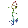
| ||||||||||||||||||
|---|---|---|---|---|---|---|---|---|---|---|---|---|---|---|---|---|---|---|---|
| 1 | 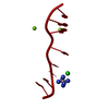
| ||||||||||||||||||
| Unit cell |
| ||||||||||||||||||
| Components on special symmetry positions |
|
-Components
| #1: DNA chain | Mass: 2460.644 Da / Num. of mol.: 1 / Source method: obtained synthetically | ||||
|---|---|---|---|---|---|
| #2: Chemical | ChemComp-CL /   Mass: 35.453 Da / Num. of mol.: 1 / Source method: obtained synthetically / Formula: Cl Mass: 35.453 Da / Num. of mol.: 1 / Source method: obtained synthetically / Formula: Cl | ||||
| #3: Chemical |   Mass: 24.305 Da / Num. of mol.: 2 / Source method: obtained synthetically / Formula: Mg Mass: 24.305 Da / Num. of mol.: 2 / Source method: obtained synthetically / Formula: Mg#4: Chemical | ChemComp-NCO / |   Mass: 161.116 Da / Num. of mol.: 1 / Source method: obtained synthetically / Formula: CoH18N6 Mass: 161.116 Da / Num. of mol.: 1 / Source method: obtained synthetically / Formula: CoH18N6#5: Water | ChemComp-HOH / |  Mass: 18.015 Da / Num. of mol.: 40 / Source method: isolated from a natural source / Formula: H2O Mass: 18.015 Da / Num. of mol.: 40 / Source method: isolated from a natural source / Formula: H2O |
-Experimental details
-Experiment
| Experiment | Method: X-RAY DIFFRACTION / Number of used crystals: 1 |
|---|
- Sample preparation
| Crystal | Density Matthews: 1.97 Å3/Da / Density % sol: 37.09 % | |||||||||||||||||||||||||||||||||||||||||||||||||||||||||||||||
|---|---|---|---|---|---|---|---|---|---|---|---|---|---|---|---|---|---|---|---|---|---|---|---|---|---|---|---|---|---|---|---|---|---|---|---|---|---|---|---|---|---|---|---|---|---|---|---|---|---|---|---|---|---|---|---|---|---|---|---|---|---|---|---|---|
| Crystal grow | Temperature: 277 K / Method: vapor diffusion, hanging drop / pH: 7 Details: magnesium chloride, spermine 4HCl, hexaamminecobalt(III) chloride, 2-methyl-2,4-pentanediol, sodium cacodylate, pH 7.0, VAPOR DIFFUSION, HANGING DROP, temperature 277K | |||||||||||||||||||||||||||||||||||||||||||||||||||||||||||||||
| Components of the solutions |
| |||||||||||||||||||||||||||||||||||||||||||||||||||||||||||||||
| Crystal grow | *PLUS Temperature: 277 K / Method: vapor diffusion, hanging drop | |||||||||||||||||||||||||||||||||||||||||||||||||||||||||||||||
| Components of the solutions | *PLUS
|
-Data collection
| Diffraction | Mean temperature: 100 K |
|---|---|
| Diffraction source | Source: SYNCHROTRON / Site: Photon Factory  / Beamline: BL-18B / Wavelength: 0.978 Å / Beamline: BL-18B / Wavelength: 0.978 Å |
| Detector | Type: ADSC QUANTUM 4 / Detector: CCD / Date: Mar 6, 2002 |
| Radiation | Protocol: SINGLE WAVELENGTH / Monochromatic (M) / Laue (L): M / Scattering type: x-ray |
| Radiation wavelength | Wavelength: 0.978 Å / Relative weight: 1 |
| Reflection | Resolution: 2.1→32 Å / Num. all: 1817 / Num. obs: 1817 / % possible obs: 100 % / Rmerge(I) obs: 0.044 / Net I/σ(I): 6.2 |
| Reflection shell | Resolution: 2.1→2.21 Å / Rmerge(I) obs: 0.393 / Mean I/σ(I) obs: 1.9 / % possible all: 100 |
| Reflection | *PLUS Num. measured all: 102876 |
| Reflection shell | *PLUS Highest resolution: 2.1 Å / % possible obs: 100 % |
- Processing
| Software |
| ||||||||||||||||||||
|---|---|---|---|---|---|---|---|---|---|---|---|---|---|---|---|---|---|---|---|---|---|
| Refinement | Method to determine structure: MOLECULAR REPLACEMENT Starting model: PDB ENTRY 1UE2 Resolution: 2.15→10 Å / σ(F): 3 Stereochemistry target values: G. PARKINSON ET AL., (1996) ACTACRYST. D52, 57-64
| ||||||||||||||||||||
| Refinement step | Cycle: LAST / Resolution: 2.15→10 Å
| ||||||||||||||||||||
| Refine LS restraints |
| ||||||||||||||||||||
| LS refinement shell | Resolution: 2.15→2.23 Å /
| ||||||||||||||||||||
| Refinement | *PLUS Lowest resolution: 10 Å / % reflection Rfree: 10 % / Rfactor Rfree: 0.239 / Rfactor Rwork: 0.227 | ||||||||||||||||||||
| Solvent computation | *PLUS | ||||||||||||||||||||
| Displacement parameters | *PLUS | ||||||||||||||||||||
| Refine LS restraints | *PLUS
|
