- PDB-1o06: Crystal structure of the Vps27p Ubiquitin Interacting Motif (UIM) -
+
Open data
ID or keywords:
Loading...
-
Basic information
Entry
Database: PDB / ID: 1o06
Title
Crystal structure of the Vps27p Ubiquitin Interacting Motif (UIM)
Components
Vacuolar protein sorting-associated protein VPS27
Keywords
TRANSPORT PROTEIN / alpha-helix / coiled-coil / tetramer
Function / homology
Function and homology information
positive regulation of protein maturation / microlipophagy / ESCRT-0 complex / microautophagy / protein retention in Golgi apparatus / protein transport to vacuole involved in ubiquitin-dependent protein catabolic process via the multivesicular body sorting pathway / ATP export / multivesicular body sorting pathway / late endosome to vacuole transport / protein targeting to vacuole ...positive regulation of protein maturation / microlipophagy / ESCRT-0 complex / microautophagy / protein retention in Golgi apparatus / protein transport to vacuole involved in ubiquitin-dependent protein catabolic process via the multivesicular body sorting pathway / ATP export / multivesicular body sorting pathway / late endosome to vacuole transport / protein targeting to vacuole / phosphatidylinositol-3-phosphate binding / ubiquitin-dependent protein catabolic process via the multivesicular body sorting pathway / cellular response to nitrogen starvation / vacuolar membrane / K48-linked polyubiquitin modification-dependent protein binding / K63-linked polyubiquitin modification-dependent protein binding / protein secretion / ubiquitin binding / endosome / endosome membrane / protein heterodimerization activity / protein domain specific binding / protein-containing complex / zinc ion binding Similarity search - Function
: / Vacuolar protein sorting-associated protein 27, GAT-like domain / Hepatocyte growth factor-regulated tyrosine kinase substrate/VPS27 / VHS domain / VHS domain / VHS domain profile. / Domain present in VPS-27, Hrs and STAM / FYVE zinc finger / FYVE zinc finger / Protein present in Fab1, YOTB, Vac1, and EEA1 ...: / Vacuolar protein sorting-associated protein 27, GAT-like domain / Hepatocyte growth factor-regulated tyrosine kinase substrate/VPS27 / VHS domain / VHS domain / VHS domain profile. / Domain present in VPS-27, Hrs and STAM / FYVE zinc finger / FYVE zinc finger / Protein present in Fab1, YOTB, Vac1, and EEA1 / Zinc finger, FYVE-related / Zinc finger FYVE/FYVE-related type profile. / ENTH/VHS / Ubiquitin interaction motif / Ubiquitin-interacting motif. / Ubiquitin interacting motif / Ubiquitin-interacting motif (UIM) domain profile. / Zinc finger, FYVE/PHD-type / Zinc finger, RING/FYVE/PHD-type Similarity search - Domain/homology
Mass: 2245.353 Da / Num. of mol.: 1 / Fragment: Residues 301-320 / Source method: obtained synthetically Details: This sequence occurs naturally in Saccharomyces cerevisiae Vps27p and was synthesized by standard peptide synthesis methods References: UniProt: P40343
In the structure databanks used in Yorodumi, some data are registered as the other names, "COVID-19 virus" and "2019-nCoV". Here are the details of the virus and the list of structure data.
Jan 31, 2019. EMDB accession codes are about to change! (news from PDBe EMDB page)
EMDB accession codes are about to change! (news from PDBe EMDB page)
The allocation of 4 digits for EMDB accession codes will soon come to an end. Whilst these codes will remain in use, new EMDB accession codes will include an additional digit and will expand incrementally as the available range of codes is exhausted. The current 4-digit format prefixed with “EMD-” (i.e. EMD-XXXX) will advance to a 5-digit format (i.e. EMD-XXXXX), and so on. It is currently estimated that the 4-digit codes will be depleted around Spring 2019, at which point the 5-digit format will come into force.
The EM Navigator/Yorodumi systems omit the EMD- prefix.
Related info.:Q: What is EMD? / ID/Accession-code notation in Yorodumi/EM Navigator
Yorodumi is a browser for structure data from EMDB, PDB, SASBDB, etc.
This page is also the successor to EM Navigator detail page, and also detail information page/front-end page for Omokage search.
The word "yorodu" (or yorozu) is an old Japanese word meaning "ten thousand". "mi" (miru) is to see.
Related info.:EMDB / PDB / SASBDB / Comparison of 3 databanks / Yorodumi Search / Aug 31, 2016. New EM Navigator & Yorodumi / Yorodumi Papers / Jmol/JSmol / Function and homology information / Changes in new EM Navigator and Yorodumi
 Movie
Movie Controller

 Yorodumi
 Open data
 Basic information
 Components
 Keywords
 Function and homology information
 X-RAY DIFFRACTION /
  Authors
 Citation
 Structure visualization
 Downloads & links
 Other downloads
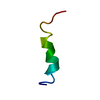

 PDBj
PDBj


 Assembly
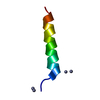


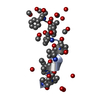
 Mass: 65.409 Da / Num. of mol.: 3 / Source method: obtained synthetically / Formula: Zn
 Mass: 18.015 Da / Num. of mol.: 26 / Source method: isolated from a natural source / Formula: H2O
 Sample preparation
 Processing