 Movie
Movie Controller

[English] 日本語
 Yorodumi
- PDB-1u9z: Crystal Structure of Phosphoribosyl Diphosphate Synthase Complexe... -
+ Open data

- Basic information
| Entry | Database: PDB / ID: 1u9z | ||||||
|---|---|---|---|---|---|---|---|
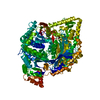
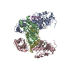
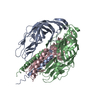
| Title | Crystal Structure of Phosphoribosyl Diphosphate Synthase Complexed with AMP and Ribose 5-Phosphate | ||||||
 Components Components | Ribose-phosphate pyrophosphokinase | ||||||
 Keywords Keywords | TRANSFERASE / PRPP synthase / ribose 5-phosphate / adenosine 5'-monophosphate | ||||||
| Function / homology |  Function and homology information Function and homology informationribose phosphate diphosphokinase complex / ribose-phosphate diphosphokinase / ribose phosphate diphosphokinase activity / 5-phosphoribose 1-diphosphate biosynthetic process / purine nucleotide biosynthetic process / kinase activity / magnesium ion binding / ATP binding / cytoplasm Similarity search - Function | ||||||
| Biological species |   Methanocaldococcus jannaschii (archaea) Methanocaldococcus jannaschii (archaea) | ||||||
| Method |  X-RAY DIFFRACTION / SYNCHROTRON / FOURIER SYNTHESIS / Resolution: 2.8 Å X-RAY DIFFRACTION / SYNCHROTRON / FOURIER SYNTHESIS / Resolution: 2.8 Å | ||||||
 Authors Authors | Kadziola, A. / Johansson, E. / Jepsen, C.H. / McGuire, J. / Larsen, S. / Hove-Jensen, B. | ||||||
 Citation Citation | Journal: J.Mol.Biol. / Year: 2005 Title: Novel class III phosphoribosyl diphosphate synthase: structure and properties of the tetrameric, phosphate-activated, non-allosterically inhibited enzyme from Methanocaldococcus jannaschii Authors: Kadziola, A. / Jepsen, C.H. / Johansson, E. / McGuire, J. / Larsen, S. / Hove-Jensen, B. | ||||||
| History |
|
- Structure visualization
| Structure viewer | Molecule: MolmilJmol/JSmol |
|---|
- Downloads & links
-Download
| PDBx/mmCIF format | 1u9z.cif.gz | 223.3 KB | Display | PDBx/mmCIF format |
|---|---|---|---|---|
| PDB format | pdb1u9z.ent.gz | 180.1 KB | Display | PDB format |
| PDBx/mmJSON format | 1u9z.json.gz | Tree view | PDBx/mmJSON format | |
| Others |  Other downloads Other downloads |
-Validation report
| Arichive directory | https://data.pdbj.org/pub/pdb/validation_reports/u9/1u9zftp://data.pdbj.org/pub/pdb/validation_reports/u9/1u9z | HTTPS FTP |
|---|
-Related structure data
| Related structure data | 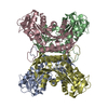 1u9ySC S: Starting model for refinement C: citing same article ( |
|---|---|
| Similar structure data |

-Links
 PDBj
PDBj


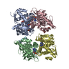
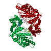
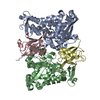
- Assembly
| Deposited unit | 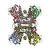
| ||||||||||
|---|---|---|---|---|---|---|---|---|---|---|---|
| 1 |
| ||||||||||
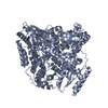
| Unit cell |
|
-Components
| #1: Protein | Mass: 31433.258 Da / Num. of mol.: 4 / Fragment: Phosphoribosyl Diphosphate Synthase Source method: isolated from a genetically manipulated source Source: (gene. exp.) Methanocaldococcus jannaschii (archaea)Gene: prs / Production host:  References: UniProt: Q58761, ribose-phosphate diphosphokinase #2: Sugar | ChemComp-R5P /   Type: saccharide / Mass: 230.110 Da / Num. of mol.: 4 / Source method: obtained synthetically / Formula: C5H11O8P Type: saccharide / Mass: 230.110 Da / Num. of mol.: 4 / Source method: obtained synthetically / Formula: C5H11O8P#3: Chemical | ChemComp-AMP /   Mass: 347.221 Da / Num. of mol.: 4 / Source method: obtained synthetically / Formula: C10H14N5O7P / Comment: AMP*YM Mass: 347.221 Da / Num. of mol.: 4 / Source method: obtained synthetically / Formula: C10H14N5O7P / Comment: AMP*YM#4: Water | ChemComp-HOH / |  Mass: 18.015 Da / Num. of mol.: 206 / Source method: isolated from a natural source / Formula: H2O Mass: 18.015 Da / Num. of mol.: 206 / Source method: isolated from a natural source / Formula: H2O |
|---|
-Experimental details
-Experiment
| Experiment | Method: X-RAY DIFFRACTION / Number of used crystals: 1 |
|---|
- Sample preparation
| Crystal | Density Matthews: 2.74 Å3/Da / Density % sol: 54.8 % |
|---|---|
| Crystal grow | Temperature: 293 K / Method: vapor diffusion, hanging drop / pH: 7 Details: PEG 4000, pH 7.0, VAPOR DIFFUSION, HANGING DROP, temperature 293K |
-Data collection
| Diffraction | Mean temperature: 100 K |
|---|---|
| Diffraction source | Source: SYNCHROTRON / Site: MAX II  / Beamline: I711 / Wavelength: 1.087 Å / Beamline: I711 / Wavelength: 1.087 Å |
| Detector | Type: MARRESEARCH / Detector: CCD / Date: Mar 11, 2003 |
| Radiation | Monochromator: single Si(111) monochromator crystal / Protocol: SINGLE WAVELENGTH / Monochromatic (M) / Laue (L): M / Scattering type: x-ray |
| Radiation wavelength | Wavelength: 1.087 Å / Relative weight: 1 |
| Reflection | Resolution: 2.8→25 Å / Num. all: 34117 / Num. obs: 34117 / % possible obs: 99.5 % / Observed criterion σ(F): 0 / Observed criterion σ(I): 0 / Rmerge(I) obs: 0.105 / Net I/σ(I): 10.5 |
| Reflection shell | Resolution: 2.8→2.87 Å / Rmerge(I) obs: 0.573 / Mean I/σ(I) obs: 1.9 / % possible all: 98.5 |
- Processing
| Software |
| |||||||||||||||||||||||||
|---|---|---|---|---|---|---|---|---|---|---|---|---|---|---|---|---|---|---|---|---|---|---|---|---|---|---|
| Refinement | Method to determine structure: FOURIER SYNTHESIS Starting model: Apo structure of Methanocaldococcus jannaschii PRPP synthase PDBID 1u9y Resolution: 2.8→25 Å Isotropic thermal model: neighbour restrained B-factor optimization Cross valid method: THROUGHOUT / σ(F): 0 / σ(I): 0 / Stereochemistry target values: Engh & Huber
| |||||||||||||||||||||||||
| Displacement parameters | Biso mean: 44.2 Å2 | |||||||||||||||||||||||||
| Refinement step | Cycle: LAST / Resolution: 2.8→25 Å
| |||||||||||||||||||||||||
| Refine LS restraints |
|
