 Movie
Movie Controller

[English] 日本語
 Yorodumi
- PDB-3s5i: Crystal structures of falcilysin, a M16 metalloprotease from the ... -
+ Open data

- Basic information
| Entry | Database: PDB / ID: 3s5i | ||||||
|---|---|---|---|---|---|---|---|
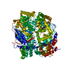
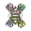
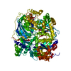
| Title | Crystal structures of falcilysin, a M16 metalloprotease from the malaria parasite Plasmodium falciparum | ||||||
 Components Components | Falcilysin | ||||||
 Keywords Keywords | HYDROLASE / M16 metalloprotease / peptidase | ||||||
| Function / homology |  Function and homology information Function and homology informationhemoglobin catabolic process / apicoplast / food vacuole / Hydrolases; Acting on peptide bonds (peptidases); Metalloendopeptidases / vacuolar membrane / protein processing / metalloendopeptidase activity / metal ion binding Similarity search - Function | ||||||
| Biological species |  | ||||||
| Method |  X-RAY DIFFRACTION / SYNCHROTRON / MOLECULAR REPLACEMENT / Resolution: 1.743 Å X-RAY DIFFRACTION / SYNCHROTRON / MOLECULAR REPLACEMENT / Resolution: 1.743 Å | ||||||
 Authors Authors | Morgunova, E. / Ponpuak, M. / Istvan, E. / Popov, A. / Goldberg, D. / Eneqvist, T. | ||||||
 Citation Citation | Journal: To be Published Title: Crystal structures of falcilysin, a M16 metalloprotease from the malaria parasite Plasmodium falciparum Authors: Morgunova, E. / Ponpuak, M. / Istvan, E. / Popov, A. / Goldberg, D. / Eneqvist, T. | ||||||
| History |
|
- Structure visualization
| Structure viewer | Molecule: MolmilJmol/JSmol |
|---|
- Downloads & links
-Download
| PDBx/mmCIF format | 3s5i.cif.gz | 467.1 KB | Display | PDBx/mmCIF format |
|---|---|---|---|---|
| PDB format | pdb3s5i.ent.gz | 378.9 KB | Display | PDB format |
| PDBx/mmJSON format | 3s5i.json.gz | Tree view | PDBx/mmJSON format | |
| Others |  Other downloads Other downloads |
-Validation report
| Arichive directory | https://data.pdbj.org/pub/pdb/validation_reports/s5/3s5iftp://data.pdbj.org/pub/pdb/validation_reports/s5/3s5i | HTTPS FTP |
|---|
-Related structure data
| Related structure data |  3s5hC  3s5kC  3s5mC  2fgeS C: citing same article ( S: Starting model for refinement |
|---|---|
| Similar structure data |

-Links
 PDBj
PDBj

- Assembly
| Deposited unit | 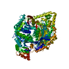
| ||||||||
|---|---|---|---|---|---|---|---|---|---|
| 1 |
| ||||||||
| Unit cell |
|
-Components
| #1: Protein | Mass: 139050.281 Da / Num. of mol.: 1 Source method: isolated from a genetically manipulated source Source: (gene. exp.) Strain: HB3 / Gene: flN, PF13_0322 / Plasmid: pET26 / Production host:  |
|---|---|
| #2: Water | ChemComp-HOH /  Mass: 18.015 Da / Num. of mol.: 1571 / Source method: isolated from a natural source / Formula: H2O Mass: 18.015 Da / Num. of mol.: 1571 / Source method: isolated from a natural source / Formula: H2O |
-Experimental details
-Experiment
| Experiment | Method: X-RAY DIFFRACTION / Number of used crystals: 1 |
|---|
- Sample preparation
| Crystal | Density Matthews: 2.33 Å3/Da / Density % sol: 47.14 % |
|---|---|
| Crystal grow | Method: vapor diffusion, sitting drop / pH: 6.2 Details: 11% PEG6000, 10%PEG400, 0.1M ADA, 20mM Mg acetate, pH 6.2, VAPOR DIFFUSION, SITTING DROP, temperature 100K |
-Data collection
| Diffraction | Mean temperature: 100 K |
|---|---|
| Diffraction source | Source: SYNCHROTRON / Site: ESRF  / Beamline: ID23-2 / Wavelength: 0.8726 Å / Beamline: ID23-2 / Wavelength: 0.8726 Å |
| Detector | Type: MARMOSAIC 225 mm CCD / Detector: CCD / Date: May 8, 2010 / Details: Pt coated Si mirror |
| Radiation | Monochromator: horizontally side diffracting Silicon 111 crystal Protocol: SINGLE WAVELENGTH / Monochromatic (M) / Laue (L): M / Scattering type: x-ray |
| Radiation wavelength | Wavelength: 0.8726 Å / Relative weight: 1 |
| Reflection | Resolution: 1.74→35.3 Å / Num. obs: 284102 / % possible obs: 99.9 % / Observed criterion σ(F): 2 / Observed criterion σ(I): 2 / Redundancy: 15.1 % / Biso Wilson estimate: 14 Å2 / Rmerge(I) obs: 0.135 / Net I/σ(I): 15 |
| Reflection shell | Resolution: 1.74→1.84 Å / Redundancy: 14.9 % / Rmerge(I) obs: 0.62 / Mean I/σ(I) obs: 4.8 / Num. unique all: 132487 / % possible all: 99.7 |
- Processing
| Software |
| |||||||||||||||||||||||||||||||||||||||||||||||||||||||||||||||||||||||||||||
|---|---|---|---|---|---|---|---|---|---|---|---|---|---|---|---|---|---|---|---|---|---|---|---|---|---|---|---|---|---|---|---|---|---|---|---|---|---|---|---|---|---|---|---|---|---|---|---|---|---|---|---|---|---|---|---|---|---|---|---|---|---|---|---|---|---|---|---|---|---|---|---|---|---|---|---|---|---|---|
| Refinement | Method to determine structure: MOLECULAR REPLACEMENT Starting model: 2FGE Resolution: 1.743→34.395 Å / SU ML: 0.19 / Isotropic thermal model: isotropic / Cross valid method: THROUGHOUT / σ(F): 1.32 / Phase error: 17.97 / Stereochemistry target values: ML
| |||||||||||||||||||||||||||||||||||||||||||||||||||||||||||||||||||||||||||||
| Solvent computation | Shrinkage radii: 0.9 Å / VDW probe radii: 1.11 Å / Solvent model: FLAT BULK SOLVENT MODEL / Bsol: 43.443 Å2 / ksol: 0.357 e/Å3 | |||||||||||||||||||||||||||||||||||||||||||||||||||||||||||||||||||||||||||||
| Displacement parameters | Biso mean: 15.8 Å2
| |||||||||||||||||||||||||||||||||||||||||||||||||||||||||||||||||||||||||||||
| Refinement step | Cycle: LAST / Resolution: 1.743→34.395 Å
| |||||||||||||||||||||||||||||||||||||||||||||||||||||||||||||||||||||||||||||
| Refine LS restraints |
| |||||||||||||||||||||||||||||||||||||||||||||||||||||||||||||||||||||||||||||
| LS refinement shell |
|
