 Movie
Movie Controller

[English] 日本語
 Yorodumi
- PDB-1u7v: Crystal Structure of the phosphorylated Smad2/Smad4 heterotrimeri... -
+ Open data

- Basic information
| Entry | Database: PDB / ID: 1u7v | ||||||
|---|---|---|---|---|---|---|---|
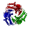
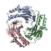
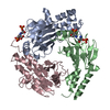
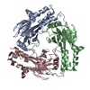
| Title | Crystal Structure of the phosphorylated Smad2/Smad4 heterotrimeric complex | ||||||
 Components Components |
| ||||||
 Keywords Keywords | SIGNALING PROTEIN / Smad / TGF-beta / signal transduction / protein complex / phosphorylation | ||||||
| Function / homology |  Function and homology information Function and homology information: / female gonad morphogenesis / negative regulation of cardiac myofibril assembly / zygotic specification of dorsal/ventral axis / metanephric mesenchyme morphogenesis / nephrogenic mesenchyme morphogenesis / somite rostral/caudal axis specification / homomeric SMAD protein complex / activin responsive factor complex / atrioventricular valve formation ...: / female gonad morphogenesis / negative regulation of cardiac myofibril assembly / zygotic specification of dorsal/ventral axis / metanephric mesenchyme morphogenesis / nephrogenic mesenchyme morphogenesis / somite rostral/caudal axis specification / homomeric SMAD protein complex / activin responsive factor complex / atrioventricular valve formation / mesendoderm development / paraxial mesoderm morphogenesis / SMAD4 MH2 Domain Mutants in Cancer / SMAD2/3 MH2 Domain Mutants in Cancer / nodal signaling pathway / sebaceous gland development / SMAD protein complex / epithelial cell migration / endoderm formation / positive regulation of luteinizing hormone secretion / formation of anatomical boundary / RUNX2 regulates bone development / pericardium development / heteromeric SMAD protein complex / filamin binding / co-SMAD binding / regulation of transforming growth factor beta2 production / positive regulation of follicle-stimulating hormone secretion / regulation of hair follicle development / RUNX3 regulates BCL2L11 (BIM) transcription / endocardial cell differentiation / epithelial to mesenchymal transition involved in endocardial cushion formation / determination of left/right asymmetry in lateral mesoderm / FOXO-mediated transcription of cell cycle genes / response to transforming growth factor beta / regulation of transforming growth factor beta receptor signaling pathway / neuron fate specification / trophoblast cell migration / secondary palate development / SMAD2/3 Phosphorylation Motif Mutants in Cancer / TGFBR1 KD Mutants in Cancer / left ventricular cardiac muscle tissue morphogenesis / odontoblast differentiation / brainstem development / positive regulation of extracellular matrix assembly / atrioventricular canal development / embryonic foregut morphogenesis / Transcriptional regulation of pluripotent stem cells / negative regulation of cardiac muscle hypertrophy / sulfate binding / cardiac conduction system development / Germ layer formation at gastrulation / primary miRNA processing / cellular response to BMP stimulus / pulmonary valve morphogenesis / transforming growth factor beta receptor binding / Signaling by BMP / Formation of definitive endoderm / SMAD protein signal transduction / embryonic cranial skeleton morphogenesis / Signaling by Activin / activin receptor signaling pathway / type I transforming growth factor beta receptor binding / Formation of axial mesoderm / positive regulation of BMP signaling pathway / Signaling by NODAL / outflow tract septum morphogenesis / pancreas development / response to cholesterol / gastrulation with mouth forming second / I-SMAD binding / cardiac muscle hypertrophy in response to stress / TGFBR3 expression / negative regulation of ossification / anterior/posterior pattern specification / aortic valve morphogenesis / Cardiogenesis / RUNX3 regulates CDKN1A transcription / endothelial cell activation / ureteric bud development / organ growth / endocardial cushion morphogenesis / insulin secretion / adrenal gland development / embryonic digit morphogenesis / branching involved in ureteric bud morphogenesis / neural crest cell differentiation / interleukin-6-mediated signaling pathway / ventricular septum morphogenesis / seminiferous tubule development / positive regulation of transforming growth factor beta receptor signaling pathway / uterus development / SMAD binding / ciliary transition zone / R-SMAD binding / TGF-beta receptor signaling activates SMADs / mesoderm formation / positive regulation of SMAD protein signal transduction / epithelial to mesenchymal transition / developmental growth Similarity search - Function | ||||||
| Biological species |  Homo sapiens (human) Homo sapiens (human) | ||||||
| Method |  X-RAY DIFFRACTION / MOLECULAR REPLACEMENT / Resolution: 2.7 Å X-RAY DIFFRACTION / MOLECULAR REPLACEMENT / Resolution: 2.7 Å | ||||||
 Authors Authors | Chacko, B.M. / Qin, B.Y. / Tiwari, A. / Shi, G. / Lam, S. / Hayward, L.J. / de Caestecker, M. / Lin, K. | ||||||
 Citation Citation | Journal: Mol.Cell / Year: 2004 Title: Structural basis of heteromeric smad protein assembly in tgf-Beta signaling Authors: Chacko, B.M. / Qin, B.Y. / Tiwari, A. / Shi, G. / Lam, S. / Hayward, L.J. / De Caestecker, M. / Lin, K. | ||||||
| History |
|
- Structure visualization
| Structure viewer | Molecule: MolmilJmol/JSmol |
|---|
- Downloads & links
-Download
| PDBx/mmCIF format | 1u7v.cif.gz | 130 KB | Display | PDBx/mmCIF format |
|---|---|---|---|---|
| PDB format | pdb1u7v.ent.gz | 102.3 KB | Display | PDB format |
| PDBx/mmJSON format | 1u7v.json.gz | Tree view | PDBx/mmJSON format | |
| Others |  Other downloads Other downloads |
-Validation report
| Arichive directory | https://data.pdbj.org/pub/pdb/validation_reports/u7/1u7vftp://data.pdbj.org/pub/pdb/validation_reports/u7/1u7v | HTTPS FTP |
|---|
-Related structure data
| Related structure data |  1u7fC  1khxS C: citing same article ( S: Starting model for refinement |
|---|---|
| Similar structure data |


-Links
 PDBj
PDBj


- Assembly
| Deposited unit | 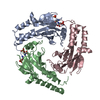
| ||||||||
|---|---|---|---|---|---|---|---|---|---|
| 1 |
| ||||||||
| Unit cell |
|
-Components
| #1: Protein | Mass: 22499.336 Da / Num. of mol.: 2 / Fragment: MH2 and Linker domains Source method: isolated from a genetically manipulated source Source: (gene. exp.) Homo sapiens (human) / Gene: SMAD2, MADH2, MADR2 / Plasmid: pTXB-1 / Production host:  #2: Protein | | Mass: 25735.330 Da / Num. of mol.: 1 / Fragment: MH2 and Linker domains Source method: isolated from a genetically manipulated source Source: (gene. exp.) Homo sapiens (human) / Gene: SMAD4, MADH4, DPC4 / Plasmid: pGEX-4T2 / Production host: Has protein modification | Y | |
|---|
-Experimental details
-Experiment
| Experiment | Method: X-RAY DIFFRACTION / Number of used crystals: 1 |
|---|
- Sample preparation
| Crystal | Density Matthews: 2.16 Å3/Da / Density % sol: 43.16 % |
|---|---|
| Crystal grow | Temperature: 298 K / Method: vapor diffusion, hanging drop / pH: 7.5 Details: 50 mM Tris-HCl, 0-15 mM magnesium chloride, 5-15% ethanol, pH 7.5, VAPOR DIFFUSION, HANGING DROP, temperature 298K |
-Data collection
| Diffraction | Mean temperature: 103 K |
|---|---|
| Diffraction source | Source: ROTATING ANODE / Type: RIGAKU RU300 / Wavelength: 1.5418 Å |
| Detector | Type: RIGAKU RAXIS IV / Detector: IMAGE PLATE / Date: Aug 10, 2002 / Details: mirrors |
| Radiation | Monochromator: Ni filter/Osmic mirror / Protocol: MAD / Monochromatic (M) / Laue (L): M / Scattering type: x-ray |
| Radiation wavelength | Wavelength: 1.5418 Å / Relative weight: 1 |
| Reflection | Resolution: 2.7→100 Å / Num. all: 17632 / Num. obs: 14546 / % possible obs: 82.5 % / Observed criterion σ(F): 0 / Observed criterion σ(I): 0 / Redundancy: 5.1 % / Rmerge(I) obs: 0.075 / Net I/σ(I): 16.7 |
| Reflection shell | Resolution: 2.7→2.8 Å / Rmerge(I) obs: 0.075 / Mean I/σ(I) obs: 3.9 / Num. unique all: 1353 / % possible all: 84.4 |
- Processing
| Software |
| |||||||||||||||||||||||||
|---|---|---|---|---|---|---|---|---|---|---|---|---|---|---|---|---|---|---|---|---|---|---|---|---|---|---|
| Refinement | Method to determine structure: MOLECULAR REPLACEMENT Starting model: PDB ENTRY 1KHX Resolution: 2.7→100 Å / Isotropic thermal model: restrained / Cross valid method: THROUGHOUT / σ(F): 0 / σ(I): 0 / Stereochemistry target values: Engh & Huber
| |||||||||||||||||||||||||
| Displacement parameters | Biso mean: 67.6 Å2 | |||||||||||||||||||||||||
| Refinement step | Cycle: LAST / Resolution: 2.7→100 Å
| |||||||||||||||||||||||||
| Refine LS restraints |
| |||||||||||||||||||||||||
| LS refinement shell | Resolution: 2.7→2.8 Å
|
