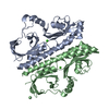
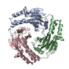
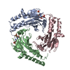
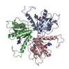
- PDB-3e4p: Crystal structure of malonate occupied DctB -
+
Open data
ID or keywords:
Loading...
-
Basic information
Entry
Database: PDB / ID: 3e4p
Title
Crystal structure of malonate occupied DctB
Components
C4-dicarboxylate transport sensor protein dctB
Keywords
TRANSFERASE / PAS DOMAIN / N-TERM HELICAL DIMERIZATION DOMAIN
Function / homology
Function and homology information
phosphorelay sensor kinase activity / histidine kinase / ATP binding / plasma membrane Similarity search - Function
Single alpha-helices involved in coiled-coils or other helix-helix interfaces - #3020 / Signal transduction histidine kinase, DctB (C4-dicarboxylate transport system regulator) / Periplasmic sensor-like domain superfamily / His Kinase A (phospho-acceptor) domain / His Kinase A (phosphoacceptor) domain / Signal transduction histidine kinase, dimerisation/phosphoacceptor domain / PAS domain / Signal transduction histidine kinase-related protein, C-terminal / Signal transduction histidine kinase, dimerisation/phosphoacceptor domain superfamily / Single alpha-helices involved in coiled-coils or other helix-helix interfaces ...Single alpha-helices involved in coiled-coils or other helix-helix interfaces - #3020 / Signal transduction histidine kinase, DctB (C4-dicarboxylate transport system regulator) / Periplasmic sensor-like domain superfamily / His Kinase A (phospho-acceptor) domain / His Kinase A (phosphoacceptor) domain / Signal transduction histidine kinase, dimerisation/phosphoacceptor domain / PAS domain / Signal transduction histidine kinase-related protein, C-terminal / Signal transduction histidine kinase, dimerisation/phosphoacceptor domain superfamily / Single alpha-helices involved in coiled-coils or other helix-helix interfaces / Histidine kinase domain / Histidine kinase domain profile. / Beta-Lactamase / Histidine kinase-, DNA gyrase B-, and HSP90-like ATPase / Helix non-globular / Histidine kinase-like ATPases / Histidine kinase/HSP90-like ATPase / Special / Histidine kinase/HSP90-like ATPase superfamily / 2-Layer Sandwich / Alpha Beta Similarity search - Domain/homology
Mass: 18.015 Da / Num. of mol.: 113 / Source method: isolated from a natural source / Formula: H2O
Sequence details
THE FEATURE OF UNIPROT (DCTB_RHIME P13633) SHOWS CONFLICT AT THE POSITION 174: N -> K (IN REF. 5). ...THE FEATURE OF UNIPROT (DCTB_RHIME P13633) SHOWS CONFLICT AT THE POSITION 174: N -> K (IN REF. 5). REFERENCE FOR THE POSITION 309 (K -> N) IS FEMS MICROBIOLOGY LETTERS 14 (1982) 95-99, DEREPRESSION OF RIBULOSE BISPHOSPHATE CARBOXYLASE ACTIVITY IN RHIZOBIUM MELILOTI SUNDARAM S. MANIAN AND FERGAL O'GARA
-
Experimental details
-
Experiment
Experiment
Method: X-RAY DIFFRACTION / Number of used crystals: 1
-
Sample preparation
Crystal
Density Matthews: 1.91 Å3/Da / Density % sol: 35.48 %
Crystal grow
Temperature: 289 K / Method: vapor diffusion / pH: 6 Details: 0.2 M SODIUM MALONATE PH 6.0, 13-15% PEG 3350, 5-10 mM STRONTIUM CHLORIDE, VAPOR DIFFUSION, temperature 289K
In the structure databanks used in Yorodumi, some data are registered as the other names, "COVID-19 virus" and "2019-nCoV". Here are the details of the virus and the list of structure data.
Jan 31, 2019. EMDB accession codes are about to change! (news from PDBe EMDB page)
EMDB accession codes are about to change! (news from PDBe EMDB page)
The allocation of 4 digits for EMDB accession codes will soon come to an end. Whilst these codes will remain in use, new EMDB accession codes will include an additional digit and will expand incrementally as the available range of codes is exhausted. The current 4-digit format prefixed with “EMD-” (i.e. EMD-XXXX) will advance to a 5-digit format (i.e. EMD-XXXXX), and so on. It is currently estimated that the 4-digit codes will be depleted around Spring 2019, at which point the 5-digit format will come into force.
The EM Navigator/Yorodumi systems omit the EMD- prefix.
Related info.:Q: What is EMD? / ID/Accession-code notation in Yorodumi/EM Navigator
Yorodumi is a browser for structure data from EMDB, PDB, SASBDB, etc.
This page is also the successor to EM Navigator detail page, and also detail information page/front-end page for Omokage search.
The word "yorodu" (or yorozu) is an old Japanese word meaning "ten thousand". "mi" (miru) is to see.
Related info.:EMDB / PDB / SASBDB / Comparison of 3 databanks / Yorodumi Search / Aug 31, 2016. New EM Navigator & Yorodumi / Yorodumi Papers / Jmol/JSmol / Function and homology information / Changes in new EM Navigator and Yorodumi
 Movie
Movie Controller


 Open data
 Basic information
 Components
 Keywords
 Function and homology information
 Sinorhizobium meliloti (bacteria)
 X-RAY DIFFRACTION /
  Authors
 Citation
 Structure visualization
 Downloads & links
 Other downloads


 PDBj
PDBj


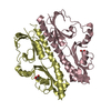
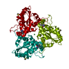
 Assembly

 Mass: 87.620 Da / Num. of mol.: 3 / Source method: obtained synthetically / Formula: Sr
 Mass: 104.061 Da / Num. of mol.: 2 / Source method: obtained synthetically / Formula: C3H4O4
 Mass: 18.015 Da / Num. of mol.: 113 / Source method: isolated from a natural source / Formula: H2O
 Sample preparation
 / Beamline: 3W1A / Wavelength: 0.979
  Processing
