 Movie
Movie Controller

[English] 日本語
 Yorodumi
- PDB-1u58: Crystal structure of the murine cytomegalovirus MHC-I homolog m144 -
+ Open data

- Basic information
| Entry | Database: PDB / ID: 1u58 | ||||||
|---|---|---|---|---|---|---|---|
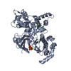
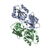
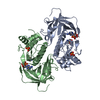
| Title | Crystal structure of the murine cytomegalovirus MHC-I homolog m144 | ||||||
 Components Components |
| ||||||
 Keywords Keywords | IMMUNE SYSTEM / MCMV / MHC-I homolog / m144 / beta-2m | ||||||
| Function / homology |  Function and homology information Function and homology informationEndosomal/Vacuolar pathway / DAP12 interactions / Antigen Presentation: Folding, assembly and peptide loading of class I MHC / ER-Phagosome pathway / DAP12 signaling / Immunoregulatory interactions between a Lymphoid and a non-Lymphoid cell / cellular defense response / Neutrophil degranulation / cellular response to iron(III) ion / negative regulation of forebrain neuron differentiation ...Endosomal/Vacuolar pathway / DAP12 interactions / Antigen Presentation: Folding, assembly and peptide loading of class I MHC / ER-Phagosome pathway / DAP12 signaling / Immunoregulatory interactions between a Lymphoid and a non-Lymphoid cell / cellular defense response / Neutrophil degranulation / cellular response to iron(III) ion / negative regulation of forebrain neuron differentiation / antigen processing and presentation of exogenous protein antigen via MHC class Ib, TAP-dependent / iron ion transport / peptide antigen assembly with MHC class I protein complex / regulation of iron ion transport / regulation of erythrocyte differentiation / HFE-transferrin receptor complex / response to molecule of bacterial origin / MHC class I peptide loading complex / T cell mediated cytotoxicity / positive regulation of T cell cytokine production / antigen processing and presentation of endogenous peptide antigen via MHC class I / MHC class I protein complex / positive regulation of receptor-mediated endocytosis / negative regulation of neurogenesis / cellular response to nicotine / positive regulation of T cell mediated cytotoxicity / multicellular organismal-level iron ion homeostasis / phagocytic vesicle membrane / negative regulation of epithelial cell proliferation / sensory perception of smell / positive regulation of cellular senescence / T cell differentiation in thymus / negative regulation of neuron projection development / protein refolding / protein homotetramerization / amyloid fibril formation / intracellular iron ion homeostasis / learning or memory / external side of plasma membrane / structural molecule activity / Golgi apparatus / protein homodimerization activity / extracellular space / membrane / cytosol Similarity search - Function | ||||||
| Biological species |  Murid herpesvirus 1 (Murine cytomegalovirus) Murid herpesvirus 1 (Murine cytomegalovirus) | ||||||
| Method |  X-RAY DIFFRACTION / SYNCHROTRON / MOLECULAR REPLACEMENT / Resolution: 1.9 Å X-RAY DIFFRACTION / SYNCHROTRON / MOLECULAR REPLACEMENT / Resolution: 1.9 Å | ||||||
 Authors Authors | Natarajan, K. / Hicks, A. / Robinson, H. / Guan, R. / Margulies, D.H. | ||||||
 Citation Citation | Journal: J.Mol.Biol. / Year: 2006 Title: Crystal structure of the murine cytomegalovirus MHC-I homolog m144. Authors: Natarajan, K. / Hicks, A. / Mans, J. / Robinson, H. / Guan, R. / Mariuzza, R.A. / Margulies, D.H. | ||||||
| History |
| ||||||
| Remark 999 | SEQUENCE THE AMINO ACID SEQUENCE OF CHAIN A IS TRANSLATED FROM THE M144 OPEN READING FRAME AS FOUND ...SEQUENCE THE AMINO ACID SEQUENCE OF CHAIN A IS TRANSLATED FROM THE M144 OPEN READING FRAME AS FOUND IN MCMV GENOME, ACCESSION NUMBER NC_004065. |
- Structure visualization
| Structure viewer | Molecule: MolmilJmol/JSmol |
|---|
- Downloads & links
-Download
| PDBx/mmCIF format | 1u58.cif.gz | 83.9 KB | Display | PDBx/mmCIF format |
|---|---|---|---|---|
| PDB format | pdb1u58.ent.gz | 62.9 KB | Display | PDB format |
| PDBx/mmJSON format | 1u58.json.gz | Tree view | PDBx/mmJSON format | |
| Others |  Other downloads Other downloads |
-Validation report
| Summary document | 1u58_validation.pdf.gz | 434 KB | Display | wwPDB validaton report |
|---|---|---|---|---|
| Full document | 1u58_full_validation.pdf.gz | 440.3 KB | Display | |
| Data in XML | 1u58_validation.xml.gz | 17.3 KB | Display | |
| Data in CIF | 1u58_validation.cif.gz | 25.4 KB | Display | |
| Arichive directory | https://data.pdbj.org/pub/pdb/validation_reports/u5/1u58ftp://data.pdbj.org/pub/pdb/validation_reports/u5/1u58 | HTTPS FTP |
-Related structure data
| Related structure data | 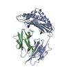 1a6zS S: Starting model for refinement |
|---|---|
| Similar structure data |


-Links
 PDBj
PDBj


- Assembly
| Deposited unit | 
| ||||||||
|---|---|---|---|---|---|---|---|---|---|
| 1 |
| ||||||||
| Unit cell |
|
-Components
| #1: Protein | Mass: 28351.463 Da / Num. of mol.: 1 Source method: isolated from a genetically manipulated source Source: (gene. exp.) Murid herpesvirus 1 (Murine cytomegalovirus)Genus: Muromegalovirus / Gene: m144 / Plasmid: pET21-b / Species (production host): Escherichia coli / Production host:  |
|---|---|
| #2: Protein | Mass: 11660.350 Da / Num. of mol.: 1 Source method: isolated from a genetically manipulated source Source: (gene. exp.) |
| #3: Water | ChemComp-HOH /  Mass: 18.015 Da / Num. of mol.: 290 / Source method: isolated from a natural source / Formula: H2O Mass: 18.015 Da / Num. of mol.: 290 / Source method: isolated from a natural source / Formula: H2O |
| Has protein modification | Y |
-Experimental details
-Experiment
| Experiment | Method: X-RAY DIFFRACTION / Number of used crystals: 1 |
|---|
- Sample preparation
| Crystal | Density Matthews: 2.96 Å3/Da / Density % sol: 58.13 % |
|---|---|
| Crystal grow | Temperature: 277 K / Method: vapor diffusion, hanging drop / pH: 6.5 Details: PEG 20K, MES, pH 6.5, VAPOR DIFFUSION, HANGING DROP, temperature 277K |
-Data collection
| Diffraction | Mean temperature: 100 K |
|---|---|
| Diffraction source | Source: SYNCHROTRON / Site: NSLS  / Beamline: X26C / Wavelength: 1 Å / Beamline: X26C / Wavelength: 1 Å |
| Detector | Type: ADSC QUANTUM 4 / Detector: CCD / Date: Jul 12, 2003 |
| Radiation | Protocol: SINGLE WAVELENGTH / Monochromatic (M) / Laue (L): M / Scattering type: x-ray |
| Radiation wavelength | Wavelength: 1 Å / Relative weight: 1 |
| Reflection | Resolution: 1.87→50 Å / Num. all: 39025 / Num. obs: 36627 / % possible obs: 98 % / Observed criterion σ(F): 2 / Observed criterion σ(I): 2 / Rmerge(I) obs: 0.11 |
| Reflection shell | Resolution: 1.87→1.94 Å / Rmerge(I) obs: 0.17 / Mean I/σ(I) obs: 4.8 / % possible all: 95.2 |
- Processing
| Software |
| ||||||||||||||||||||
|---|---|---|---|---|---|---|---|---|---|---|---|---|---|---|---|---|---|---|---|---|---|
| Refinement | Method to determine structure: MOLECULAR REPLACEMENT Starting model: PDB ENTRY 1A6Z Resolution: 1.9→41.14 Å / Isotropic thermal model: anisotropic / Cross valid method: THROUGHOUT / σ(F): 0 / σ(I): 0 / Stereochemistry target values: Engh & Huber
| ||||||||||||||||||||
| Displacement parameters |
| ||||||||||||||||||||
| Refine analyze | Luzzati coordinate error obs: 0.22 Å / Luzzati d res low obs: 5 Å / Luzzati sigma a obs: 0.17 Å | ||||||||||||||||||||
| Refinement step | Cycle: LAST / Resolution: 1.9→41.14 Å
| ||||||||||||||||||||
| Refine LS restraints |
| ||||||||||||||||||||
| LS refinement shell | Resolution: 1.9→2.02 Å / Rfactor Rfree error: 0.012
|
