 Movie
Movie Controller


+ Open data

- Basic information
| Entry | Database: PDB / ID: 1a6z | ||||||
|---|---|---|---|---|---|---|---|
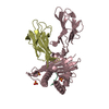
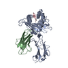
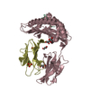
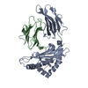
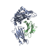
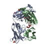
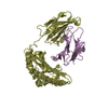
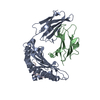
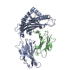
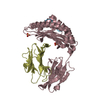
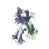
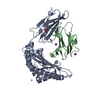
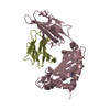
| Title | HFE (HUMAN) HEMOCHROMATOSIS PROTEIN | ||||||
 Components Components |
| ||||||
 Keywords Keywords | MHC CLASS I COMPLEX / HFE / HEREDITARY HEMOCHROMATOSIS / MHC CLASS I | ||||||
| Function / homology |  Function and homology information Function and homology informationnegative regulation of antigen processing and presentation of endogenous peptide antigen via MHC class I / response to iron ion starvation / negative regulation of T cell cytokine production / negative regulation of CD8-positive, alpha-beta T cell activation / co-receptor binding / transferrin receptor binding / regulation of protein localization to cell surface / Transferrin endocytosis and recycling / basal part of cell / response to iron ion ...negative regulation of antigen processing and presentation of endogenous peptide antigen via MHC class I / response to iron ion starvation / negative regulation of T cell cytokine production / negative regulation of CD8-positive, alpha-beta T cell activation / co-receptor binding / transferrin receptor binding / regulation of protein localization to cell surface / Transferrin endocytosis and recycling / basal part of cell / response to iron ion / positive regulation of peptide hormone secretion / positive regulation of SMAD protein signal transduction / beta-2-microglobulin binding / BMP signaling pathway / transporter activator activity / negative regulation of ubiquitin-dependent protein catabolic process / negative regulation of proteasomal ubiquitin-dependent protein catabolic process / receptor-mediated endocytosis / early endosome lumen / Nef mediated downregulation of MHC class I complex cell surface expression / DAP12 interactions / Endosomal/Vacuolar pathway / T cell mediated cytotoxicity / Antigen Presentation: Folding, assembly and peptide loading of class I MHC / regulation of iron ion transport / cellular response to iron(III) ion / negative regulation of iron ion transport / negative regulation of forebrain neuron differentiation / antigen processing and presentation of exogenous protein antigen via MHC class Ib, TAP-dependent / peptide antigen assembly with MHC class I protein complex / ER to Golgi transport vesicle membrane / regulation of erythrocyte differentiation / response to molecule of bacterial origin / HFE-transferrin receptor complex / MHC class I peptide loading complex / transferrin transport / cellular response to iron ion / negative regulation of receptor-mediated endocytosis / positive regulation of T cell cytokine production / antigen processing and presentation of endogenous peptide antigen via MHC class I / MHC class I protein complex / peptide antigen assembly with MHC class II protein complex / negative regulation of neurogenesis / MHC class II protein complex / cellular response to nicotine / positive regulation of receptor-mediated endocytosis / multicellular organismal-level iron ion homeostasis / positive regulation of T cell mediated cytotoxicity / recycling endosome / specific granule lumen / antigen processing and presentation of exogenous peptide antigen via MHC class II / positive regulation of immune response / peptide antigen binding / phagocytic vesicle membrane / recycling endosome membrane / positive regulation of T cell activation / negative regulation of epithelial cell proliferation / Interferon gamma signaling / Immunoregulatory interactions between a Lymphoid and a non-Lymphoid cell / apical part of cell / sensory perception of smell / Modulation by Mtb of host immune system / positive regulation of cellular senescence / tertiary granule lumen / MHC class II protein complex binding / T cell differentiation in thymus / DAP12 signaling / late endosome membrane / negative regulation of neuron projection development / protein refolding / ER-Phagosome pathway / protein-containing complex assembly / cytoplasmic vesicle / early endosome membrane / amyloid fibril formation / protein homotetramerization / intracellular iron ion homeostasis / learning or memory / early endosome / cell surface receptor signaling pathway / endoplasmic reticulum lumen / Amyloid fiber formation / signaling receptor binding / Golgi membrane / external side of plasma membrane / lysosomal membrane / focal adhesion / Neutrophil degranulation / positive regulation of gene expression / perinuclear region of cytoplasm / SARS-CoV-2 activates/modulates innate and adaptive immune responses / structural molecule activity / Golgi apparatus / endoplasmic reticulum / protein homodimerization activity / : / extracellular exosome / extracellular region / membrane / identical protein binding Similarity search - Function | ||||||
| Biological species |  Homo sapiens (human) Homo sapiens (human) | ||||||
| Method |  X-RAY DIFFRACTION / SYNCHROTRON / MOLECULAR REPLACEMENT / Resolution: 2.6 Å X-RAY DIFFRACTION / SYNCHROTRON / MOLECULAR REPLACEMENT / Resolution: 2.6 Å | ||||||
 Authors Authors | Lebron, J.A. / Bennett, M.J. / Vaughn, D.E. / Chirino, A.J. / Snow, P.M. / Mintier, G.A. / Feder, J.N. / Bjorkman, P.J. | ||||||
 Citation Citation | Journal: Cell(Cambridge,Mass.) / Year: 1998 Title: Crystal structure of the hemochromatosis protein HFE and characterization of its interaction with transferrin receptor. Authors: Lebron, J.A. / Bennett, M.J. / Vaughn, D.E. / Chirino, A.J. / Snow, P.M. / Mintier, G.A. / Feder, J.N. / Bjorkman, P.J. | ||||||
| History |
|
- Structure visualization
| Structure viewer | Molecule: MolmilJmol/JSmol |
|---|
- Downloads & links
-Download
| PDBx/mmCIF format | 1a6z.cif.gz | 160 KB | Display | PDBx/mmCIF format |
|---|---|---|---|---|
| PDB format | pdb1a6z.ent.gz | 127.5 KB | Display | PDB format |
| PDBx/mmJSON format | 1a6z.json.gz | Tree view | PDBx/mmJSON format | |
| Others |  Other downloads Other downloads |
-Validation report
| Arichive directory | https://data.pdbj.org/pub/pdb/validation_reports/a6/1a6zftp://data.pdbj.org/pub/pdb/validation_reports/a6/1a6z | HTTPS FTP |
|---|
-Related structure data
| Related structure data | 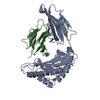 2clrS S: Starting model for refinement |
|---|---|
| Similar structure data |


-Links
 PDBj
PDBj


- Assembly
| Deposited unit | 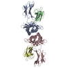
| ||||||||||||
|---|---|---|---|---|---|---|---|---|---|---|---|---|---|
| 1 | 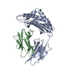
| ||||||||||||
| 2 | 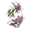
| ||||||||||||
| Unit cell |
| ||||||||||||
| Noncrystallographic symmetry (NCS) | NCS oper:
|
-Components
| #1: Protein | Mass: 32339.434 Da / Num. of mol.: 2 Source method: isolated from a genetically manipulated source Source: (gene. exp.) Homo sapiens (human) / Gene: HFE / Organ: OVARY / Cell line (production host): CHO / Cellular location (production host): SECRETED / Production host:   Cricetulus griseus (Chinese hamster) / References: UniProt: Q30201 Cricetulus griseus (Chinese hamster) / References: UniProt: Q30201#2: Protein | Mass: 11748.160 Da / Num. of mol.: 2 / Source method: isolated from a natural source / Source: (natural) Homo sapiens (human) / Organ: OVARY / References: UniProt: P61769#3: Water | ChemComp-HOH / |  Mass: 18.015 Da / Num. of mol.: 23 / Source method: isolated from a natural source / Formula: H2O Mass: 18.015 Da / Num. of mol.: 23 / Source method: isolated from a natural source / Formula: H2OHas protein modification | Y | |
|---|
-Experimental details
-Experiment
| Experiment | Method: X-RAY DIFFRACTION / Number of used crystals: 1 |
|---|
- Sample preparation
| Crystal | Density Matthews: 2.89 Å3/Da / Density % sol: 57 % | |||||||||||||||||||||||||
|---|---|---|---|---|---|---|---|---|---|---|---|---|---|---|---|---|---|---|---|---|---|---|---|---|---|---|
| Crystal grow | pH: 6.5 / Details: pH 6.5 | |||||||||||||||||||||||||
| Crystal grow | *PLUS pH: 5.9 / Method: vapor diffusion, hanging dropDetails: drop contained 1:1 mixture of protein and reservoir solution | |||||||||||||||||||||||||
| Components of the solutions | *PLUS
|
-Data collection
| Diffraction | Mean temperature: 123 K |
|---|---|
| Diffraction source | Source: SYNCHROTRON / Site: NSLS  / Beamline: X4A / Wavelength: 1.07 / Beamline: X4A / Wavelength: 1.07 |
| Detector | Type: FUJI / Detector: IMAGE PLATE / Date: Jul 1, 1997 / Details: MIRRORS |
| Radiation | Monochromator: SI(111) / Monochromatic (M) / Laue (L): M / Scattering type: x-ray |
| Radiation wavelength | Wavelength: 1.07 Å / Relative weight: 1 |
| Reflection | Resolution: 2.6→15 Å / Num. obs: 187780 / % possible obs: 98 % / Observed criterion σ(I): -3 / Redundancy: 5 % / Biso Wilson estimate: 67.7 Å2 / Rmerge(I) obs: 0.059 / Net I/σ(I): 24 |
| Reflection shell | Resolution: 2.6→2.7 Å / Redundancy: 3.5 % / Rmerge(I) obs: 0.421 / Mean I/σ(I) obs: 3 / % possible all: 97 |
| Reflection | *PLUS Num. obs: 32145 / Num. measured all: 188780 |
| Reflection shell | *PLUS % possible obs: 97 % |
- Processing
| Software |
| ||||||||||||||||||||||||||||||||||||||||||||||||||||||||||||||||||||||||||||||||
|---|---|---|---|---|---|---|---|---|---|---|---|---|---|---|---|---|---|---|---|---|---|---|---|---|---|---|---|---|---|---|---|---|---|---|---|---|---|---|---|---|---|---|---|---|---|---|---|---|---|---|---|---|---|---|---|---|---|---|---|---|---|---|---|---|---|---|---|---|---|---|---|---|---|---|---|---|---|---|---|---|---|
| Refinement | Method to determine structure: MOLECULAR REPLACEMENT Starting model: PDB ENTRY 2CLR Resolution: 2.6→15 Å / Rfactor Rfree error: 0.007 / Data cutoff high absF: 10000000 / Data cutoff low absF: 0.001 / Isotropic thermal model: RESTRAINED / Cross valid method: THROUGHOUT / σ(F): 0 / Details: BULK SOLVENT MODEL USED
| ||||||||||||||||||||||||||||||||||||||||||||||||||||||||||||||||||||||||||||||||
| Displacement parameters | Biso mean: 63.2 Å2
| ||||||||||||||||||||||||||||||||||||||||||||||||||||||||||||||||||||||||||||||||
| Refinement step | Cycle: LAST / Resolution: 2.6→15 Å
| ||||||||||||||||||||||||||||||||||||||||||||||||||||||||||||||||||||||||||||||||
| Refine LS restraints |
| ||||||||||||||||||||||||||||||||||||||||||||||||||||||||||||||||||||||||||||||||
| Refine LS restraints NCS | NCS model details: RESTRAINED / Rms dev Biso : 7.4 Å2 / Rms dev position: 0.02 Å / Weight Biso : 2 / Weight position: 300 | ||||||||||||||||||||||||||||||||||||||||||||||||||||||||||||||||||||||||||||||||
| LS refinement shell | Resolution: 2.6→2.71 Å / Rfactor Rfree error: 0.03 / Total num. of bins used: 8
| ||||||||||||||||||||||||||||||||||||||||||||||||||||||||||||||||||||||||||||||||
| Xplor file |
| ||||||||||||||||||||||||||||||||||||||||||||||||||||||||||||||||||||||||||||||||
| Software | *PLUS Name: X-PLOR / Version: 3.857 / Classification: refinement | ||||||||||||||||||||||||||||||||||||||||||||||||||||||||||||||||||||||||||||||||
| Refinement | *PLUS | ||||||||||||||||||||||||||||||||||||||||||||||||||||||||||||||||||||||||||||||||
| Solvent computation | *PLUS | ||||||||||||||||||||||||||||||||||||||||||||||||||||||||||||||||||||||||||||||||
| Displacement parameters | *PLUS | ||||||||||||||||||||||||||||||||||||||||||||||||||||||||||||||||||||||||||||||||
| Refine LS restraints | *PLUS
| ||||||||||||||||||||||||||||||||||||||||||||||||||||||||||||||||||||||||||||||||
| LS refinement shell | *PLUS Rfactor obs: 0.399 |
