 Movie
Movie Controller

[English] 日本語
 Yorodumi
- PDB-1tvn: Cellulase cel5G from Pseudoalteromonas haloplanktis, A family GH ... -
+ Open data

- Basic information
| Entry | Database: PDB / ID: 1tvn | ||||||
|---|---|---|---|---|---|---|---|
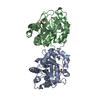
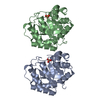
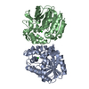
| Title | Cellulase cel5G from Pseudoalteromonas haloplanktis, A family GH 5-2 enzyme | ||||||
 Components Components | cellulase | ||||||
 Keywords Keywords | HYDROLASE / Glycoside hydrolase / Clan GH-A / Family 5-2 / Cellulase | ||||||
| Function / homology |  Function and homology information Function and homology informationcellulase / cellulase activity / polysaccharide catabolic process / carbohydrate binding / cell adhesion / calcium ion binding / extracellular region Similarity search - Function | ||||||
| Biological species |  Pseudoalteromonas haloplanktis (bacteria) Pseudoalteromonas haloplanktis (bacteria) | ||||||
| Method |  X-RAY DIFFRACTION / SYNCHROTRON / MOLECULAR REPLACEMENT / Resolution: 1.41 Å X-RAY DIFFRACTION / SYNCHROTRON / MOLECULAR REPLACEMENT / Resolution: 1.41 Å | ||||||
 Authors Authors | Violot, S. / Haser, R. / Aghajari, N. | ||||||
 Citation Citation | Journal: J.Mol.Biol. / Year: 2005 Title: Structure of a Full Length Psychrophilic Cellulase from Pseudoalteromonas haloplanktis revealed by X-ray Diffraction and Small Angle X-ray Scattering Authors: Violot, S. / Aghajari, N. / Czjzek, M. / Feller, G. / Sonan, G.K. / Gouet, P. / Gerday, C. / Haser, R. / Receveur-Brechot, V. #1: Journal: Acta Crystallogr.,Sect.D / Year: 2003 Title: Expression, purification, crystallization and preliminary X-ray crystallographic studies of a psychrophilic cellulase from Pseudoalteromonas haloplanktis Authors: Violot, S. / Haser, R. / Sonan, G. / Georlette, D. / Feller, G. / Aghajari, N. | ||||||
| History |
|
- Structure visualization
| Structure viewer | Molecule: MolmilJmol/JSmol |
|---|
- Downloads & links
-Download
| PDBx/mmCIF format | 1tvn.cif.gz | 138.1 KB | Display | PDBx/mmCIF format |
|---|---|---|---|---|
| PDB format | pdb1tvn.ent.gz | 108.5 KB | Display | PDB format |
| PDBx/mmJSON format | 1tvn.json.gz | Tree view | PDBx/mmJSON format | |
| Others |  Other downloads Other downloads |
-Validation report
| Arichive directory | https://data.pdbj.org/pub/pdb/validation_reports/tv/1tvnftp://data.pdbj.org/pub/pdb/validation_reports/tv/1tvn | HTTPS FTP |
|---|
-Related structure data
| Related structure data | 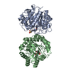 1tvpC 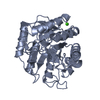 1egzS C: citing same article ( S: Starting model for refinement |
|---|---|
| Similar structure data |


-Links
 PDBj
PDBj- Assembly
| Deposited unit | 
| ||||||||
|---|---|---|---|---|---|---|---|---|---|
| 1 |
| ||||||||
| Unit cell |
|
-Components
| #1: Protein | Mass: 31914.918 Da / Num. of mol.: 2 / Fragment: CATALYTIC DOMAIN Source method: isolated from a genetically manipulated source Source: (gene. exp.) Pseudoalteromonas haloplanktis (bacteria)Gene: celG / Plasmid: pET22b / Production host: #2: Water | ChemComp-HOH / |  Mass: 18.015 Da / Num. of mol.: 776 / Source method: isolated from a natural source / Formula: H2O Mass: 18.015 Da / Num. of mol.: 776 / Source method: isolated from a natural source / Formula: H2O |
|---|
-Experimental details
-Experiment
| Experiment | Method: X-RAY DIFFRACTION / Number of used crystals: 1 |
|---|
- Sample preparation
| Crystal | Density Matthews: 1.8 Å3/Da / Density % sol: 33 % |
|---|---|
| Crystal grow | Temperature: 292 K / Method: vapor diffusion, hanging drop / pH: 7.5 Details: 1.3M tri-sodium citrate dihydrate, 40% (v/v) glycerol, 0.1M HEPES, pH 7.5, VAPOR DIFFUSION, HANGING DROP, temperature 292K |
-Data collection
| Diffraction | Mean temperature: 100 K |
|---|---|
| Diffraction source | Source: SYNCHROTRON / Site: ESRF  / Beamline: BM14 / Wavelength: 0.9559 Å / Beamline: BM14 / Wavelength: 0.9559 Å |
| Detector | Type: MARRESEARCH / Detector: CCD / Date: Aug 30, 2001 / Details: mirrors |
| Radiation | Monochromator: Si 111 CHANNEL / Protocol: SINGLE WAVELENGTH / Monochromatic (M) / Laue (L): M / Scattering type: x-ray |
| Radiation wavelength | Wavelength: 0.9559 Å / Relative weight: 1 |
| Reflection | Resolution: 1.41→14.008 Å / Num. all: 209086 / Num. obs: 87830 / % possible obs: 95.1 % / Observed criterion σ(F): 3 / Observed criterion σ(I): 3 / Redundancy: 2.4 % / Biso Wilson estimate: 11.8 Å2 / Rsym value: 0.043 |
| Reflection shell | Resolution: 1.41→1.48 Å / Redundancy: 1.8 % / Num. unique all: 9973 / Rsym value: 0.175 / % possible all: 75.3 |
- Processing
| Software |
| ||||||||||||||||||||||||||||||||||||
|---|---|---|---|---|---|---|---|---|---|---|---|---|---|---|---|---|---|---|---|---|---|---|---|---|---|---|---|---|---|---|---|---|---|---|---|---|---|
| Refinement | Method to determine structure: MOLECULAR REPLACEMENT Starting model: PDB ENTRY 1EGZ Resolution: 1.41→13.76 Å / Rfactor Rfree error: 0.003 / Data cutoff high absF: 1740239.82 / Data cutoff low absF: 0 / Isotropic thermal model: RESTRAINED / Cross valid method: THROUGHOUT / σ(F): 0 / σ(I): 0 / Stereochemistry target values: Engh & Huber
| ||||||||||||||||||||||||||||||||||||
| Solvent computation | Solvent model: FLAT MODEL / Bsol: 26.2331 Å2 / ksol: 0.392917 e/Å3 | ||||||||||||||||||||||||||||||||||||
| Displacement parameters | Biso mean: 11.1 Å2
| ||||||||||||||||||||||||||||||||||||
| Refine analyze |
| ||||||||||||||||||||||||||||||||||||
| Refinement step | Cycle: LAST / Resolution: 1.41→13.76 Å
| ||||||||||||||||||||||||||||||||||||
| Refine LS restraints |
| ||||||||||||||||||||||||||||||||||||
| LS refinement shell | Resolution: 1.41→1.49 Å / Rfactor Rfree error: 0.01 / Total num. of bins used: 6
| ||||||||||||||||||||||||||||||||||||
| Xplor file |
|
