 Movie
Movie Controller


+ Open data

- Basic information
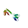
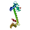
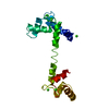
| Entry | Database: PDB / ID: 1tn4 | ||||||
|---|---|---|---|---|---|---|---|
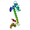
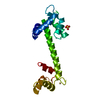
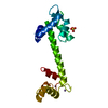
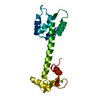
| Title | FOUR CALCIUM TNC | ||||||
 Components Components | TROPONIN C | ||||||
 Keywords Keywords | CONTRACTILE SYSTEM PROTEIN / CALCIUM REGULATION / CALMODULIN SUPERFAMILY | ||||||
| Function / homology |  Function and homology information Function and homology information | ||||||
| Biological species |  | ||||||
| Method |  X-RAY DIFFRACTION / SINGLE ISOMORPHOUS REPLACEMENT, DENSITY MODIFICATION / Resolution: 1.95 Å X-RAY DIFFRACTION / SINGLE ISOMORPHOUS REPLACEMENT, DENSITY MODIFICATION / Resolution: 1.95 Å | ||||||
 Authors Authors | Love, M.L. / Dominguez, R. / Houdusse, A. / Cohen, C. | ||||||
 Citation Citation | Journal: Structure / Year: 1997 Title: Structures of four Ca2+-bound troponin C at 2.0 A resolution: further insights into the Ca2+-switch in the calmodulin superfamily. Authors: Houdusse, A. / Love, M.L. / Dominguez, R. / Grabarek, Z. / Cohen, C. | ||||||
| History |
|
- Structure visualization
| Structure viewer | Molecule: MolmilJmol/JSmol |
|---|
- Downloads & links
-Download
| PDBx/mmCIF format | 1tn4.cif.gz | 49.8 KB | Display | PDBx/mmCIF format |
|---|---|---|---|---|
| PDB format | pdb1tn4.ent.gz | 34.3 KB | Display | PDB format |
| PDBx/mmJSON format | 1tn4.json.gz | Tree view | PDBx/mmJSON format | |
| Others |  Other downloads Other downloads |
-Validation report
| Arichive directory | https://data.pdbj.org/pub/pdb/validation_reports/tn/1tn4ftp://data.pdbj.org/pub/pdb/validation_reports/tn/1tn4 | HTTPS FTP |
|---|
-Related structure data


-Links
 PDBj
PDBj
- Assembly
| Deposited unit | 
| ||||||||
|---|---|---|---|---|---|---|---|---|---|
| 1 |
| ||||||||
| Unit cell |
|
-Components
| #1: Protein | Mass: 17991.801 Da / Num. of mol.: 1 / Mutation: C98L Source method: isolated from a genetically manipulated source Details: RABBIT SKELETAL TROPONIN C / Source: (gene. exp.)  | ||
|---|---|---|---|
| #2: Chemical | ChemComp-CA /   Mass: 40.078 Da / Num. of mol.: 7 / Source method: obtained synthetically / Formula: Ca Mass: 40.078 Da / Num. of mol.: 7 / Source method: obtained synthetically / Formula: Ca#3: Water | ChemComp-HOH / |  Mass: 18.015 Da / Num. of mol.: 181 / Source method: isolated from a natural source / Formula: H2O Mass: 18.015 Da / Num. of mol.: 181 / Source method: isolated from a natural source / Formula: H2O |
-Experimental details
-Experiment
| Experiment | Method: X-RAY DIFFRACTION / Number of used crystals: 2 |
|---|
- Sample preparation
| Crystal | Density Matthews: 2.6 Å3/Da / Density % sol: 53 % | ||||||||||||||||||||||||
|---|---|---|---|---|---|---|---|---|---|---|---|---|---|---|---|---|---|---|---|---|---|---|---|---|---|
| Crystal grow | pH: 7.2 Details: 54% MPD 50 MM HEPES, PH 7.2 10 MM CACL2 1 MM NA-AZIDE | ||||||||||||||||||||||||
| Crystal | *PLUS | ||||||||||||||||||||||||
| Crystal grow | *PLUS Temperature: 4 ℃ / Method: vapor diffusion / Details: used to seeding | ||||||||||||||||||||||||
| Components of the solutions | *PLUS
|
-Data collection
| Diffraction | Mean temperature: 160 K |
|---|---|
| Diffraction source | Source: ROTATING ANODE / Type: ELLIOTT GX-13 / Wavelength: 1.5418 |
| Detector | Type: MARRESEARCH / Detector: IMAGE PLATE / Date: Jun 25, 1996 / Details: MIRRORS |
| Radiation | Monochromator: NI FILTER / Monochromatic (M) / Laue (L): M / Scattering type: x-ray |
| Radiation wavelength | Wavelength: 1.5418 Å / Relative weight: 1 |
| Reflection | Resolution: 1.93→25 Å / Num. obs: 14374 / % possible obs: 97.8 % / Redundancy: 11 % / Rmerge(I) obs: 0.072 / Rsym value: 0.09 / Net I/σ(I): 10.8 |
| Reflection shell | Resolution: 1.9→2.2 Å / Redundancy: 6.1 % / Rmerge(I) obs: 0.16 / Mean I/σ(I) obs: 24 / Rsym value: 0.16 / % possible all: 92.05 |
| Reflection | *PLUS Num. measured all: 157890 |
- Processing
| Software |
| ||||||||||||||||||||||||||||||||||||||||||||||||||||||||||||
|---|---|---|---|---|---|---|---|---|---|---|---|---|---|---|---|---|---|---|---|---|---|---|---|---|---|---|---|---|---|---|---|---|---|---|---|---|---|---|---|---|---|---|---|---|---|---|---|---|---|---|---|---|---|---|---|---|---|---|---|---|---|
| Refinement | Method to determine structure: SINGLE ISOMORPHOUS REPLACEMENT, DENSITY MODIFICATION Resolution: 1.95→10 Å / Cross valid method: A POSTERIORI / Details: X-PLOR (BRUNGER) ALSO WAS USED.
| ||||||||||||||||||||||||||||||||||||||||||||||||||||||||||||
| Refinement step | Cycle: LAST / Resolution: 1.95→10 Å
| ||||||||||||||||||||||||||||||||||||||||||||||||||||||||||||
| Refine LS restraints |
| ||||||||||||||||||||||||||||||||||||||||||||||||||||||||||||
| Software | *PLUS Name: ARP / Classification: refinement | ||||||||||||||||||||||||||||||||||||||||||||||||||||||||||||
| Refinement | *PLUS Rfactor obs: 0.189 | ||||||||||||||||||||||||||||||||||||||||||||||||||||||||||||
| Solvent computation | *PLUS | ||||||||||||||||||||||||||||||||||||||||||||||||||||||||||||
| Displacement parameters | *PLUS | ||||||||||||||||||||||||||||||||||||||||||||||||||||||||||||
| Refine LS restraints | *PLUS
|
