 Movie
Movie Controller

[English] 日本語
 Yorodumi
- PDB-5tnc: REFINED CRYSTAL STRUCTURE OF TROPONIN C FROM TURKEY SKELETAL MUSC... -
+ Open data

- Basic information
| Entry | Database: PDB / ID: 5tnc | |||||||||
|---|---|---|---|---|---|---|---|---|---|---|
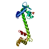
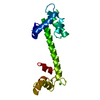
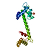
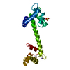
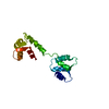
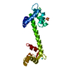
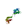
| Title | REFINED CRYSTAL STRUCTURE OF TROPONIN C FROM TURKEY SKELETAL MUSCLE AT 2.0 ANGSTROMS RESOLUTION | |||||||||
 Components Components | TROPONIN-C | |||||||||
 Keywords Keywords | CONTRACTILE SYSTEM PROTEINS | |||||||||
| Function / homology |  Function and homology information Function and homology information | |||||||||
| Biological species |  | |||||||||
| Method |  X-RAY DIFFRACTION / Resolution: 2 Å X-RAY DIFFRACTION / Resolution: 2 Å | |||||||||
 Authors Authors | Herzberg, O. / James, M.N.G. | |||||||||
 Citation Citation | Journal: J.Mol.Biol. / Year: 1988 Title: Refined crystal structure of troponin C from turkey skeletal muscle at 2.0 A resolution. Authors: Herzberg, O. / James, M.N. #1: Journal: Methods Enzymol. / Year: 1987Title: Molecular Structure of Troponin C and its Implications for the Ca2+ Triggering of Muscle Contraction Authors: Herzberg, O. / Moult, J. / James, M.N.G. #2: Journal: Calcium-Binding Proteins in Health and Disease / Year: 1987Title: Conformational Flexibility of Troponin C Authors: Herzberg, O. / Moult, J. / James, M.N.G. #3: Journal: Ciba Found.Symp. / Year: 1986Title: Calcium Binding to Skeletal Muscle Troponin C and the Regulation of Muscle Contraction Authors: Herzberg, O. / Moult, J. / James, M.N.G. #4: Journal: FEBS Lett. / Year: 1986Title: Crystallographic Determination of Lanthanide Ion Binding to Troponin C Authors: Herzberg, O. / James, M.N.G. #5: Journal: J.Biol.Chem. / Year: 1986Title: A Model for the Ca2+-Induced Conformational Transition of Troponin C. A Trigger for Muscle Contraction Authors: Herzberg, O. / Moult, J. / James, M.N.G. #6: Journal: Biochemistry / Year: 1985Title: Common Structural Framework of the Two Ca2+(Slash)Mg2+ Binding Loops of Troponin C and Other Ca2+ Binding Proteins Authors: Herzberg, O. / James, M.N.G. #7: Journal: Nature / Year: 1985Title: Structure of the Calcium Regulatory Muscle Protein Troponin-C at 2.8 Angstroms Resolution Authors: Herzberg, O. / James, M.N.G. #8: Journal: J.Mol.Biol. / Year: 1984Title: Crystallographic Data for Troponin C from Turkey Skeletal Muscle Authors: Herzberg, O. / Hayakawa, K. / James, M.N.G. #9: Journal: Acta Crystallogr.,Sect.A / Year: 1984Title: The Crystal Structure of Troponin C from Turkey Skeletal Muscle at 2.8 Angstroms Resolution Authors: Herzberg, O. / James, M.N.G. | |||||||||
| History |
|
- Structure visualization
| Structure viewer | Molecule: MolmilJmol/JSmol |
|---|
- Downloads & links
-Download
| PDBx/mmCIF format | 5tnc.cif.gz | 53.8 KB | Display | PDBx/mmCIF format |
|---|---|---|---|---|
| PDB format | pdb5tnc.ent.gz | 34.2 KB | Display | PDB format |
| PDBx/mmJSON format | 5tnc.json.gz | Tree view | PDBx/mmJSON format | |
| Others |  Other downloads Other downloads |
-Validation report
| Arichive directory | https://data.pdbj.org/pub/pdb/validation_reports/tn/5tncftp://data.pdbj.org/pub/pdb/validation_reports/tn/5tnc | HTTPS FTP |
|---|
-Related structure data
| Similar structure data |
|---|

-Links
 PDBj
PDBj

- Assembly
| Deposited unit | 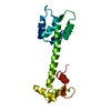
| ||||||||
|---|---|---|---|---|---|---|---|---|---|
| 1 |
| ||||||||
| Unit cell |
| ||||||||
| Atom site foot note | 1: SEE REMARK 4. / 2: SEE REMARK 6. |
-Components
| #1: Protein | Mass: 18320.209 Da / Num. of mol.: 1 Source method: isolated from a genetically manipulated source Source: (gene. exp.) | ||||||
|---|---|---|---|---|---|---|---|
| #2: Chemical |   Mass: 40.078 Da / Num. of mol.: 2 / Source method: obtained synthetically / Formula: Ca Mass: 40.078 Da / Num. of mol.: 2 / Source method: obtained synthetically / Formula: Ca#3: Water | ChemComp-HOH / |  Mass: 18.015 Da / Num. of mol.: 157 / Source method: isolated from a natural source / Formula: H2O Mass: 18.015 Da / Num. of mol.: 157 / Source method: isolated from a natural source / Formula: H2ONonpolymer details | THE WATER MOLECULES ARE ARRANGED IN DESCENDING ORDER OF RELIABILITY WHERE RELIABILITY IS DEFINED BY ...THE WATER MOLECULES ARE ARRANGED IN DESCENDING | Sequence details | BASED ON AMINO ACID COMPOSITION AND THE ELECTRON DENSITY MAP, ALA 99 IN CHICKEN TNC WAS IDENTIFIED ...BASED ON AMINO ACID COMPOSITIO | |
-Experimental details
-Experiment
| Experiment | Method: X-RAY DIFFRACTION |
|---|
- Sample preparation
| Crystal | Density Matthews: 2.13 Å3/Da / Density % sol: 42.12 % | ||||||||||||||||||||||||||||||
|---|---|---|---|---|---|---|---|---|---|---|---|---|---|---|---|---|---|---|---|---|---|---|---|---|---|---|---|---|---|---|---|
| Crystal grow | *PLUS Method: unknown / PH range low: 5 / PH range high: 4.9 | ||||||||||||||||||||||||||||||
| Components of the solutions | *PLUS
|
-Data collection
| Reflection | *PLUS Highest resolution: 2 Å / Num. obs: 8054 / Observed criterion σ(I): 2 / Rmerge(I) obs: 0.06 / Num. measured all: 10825 |
|---|
- Processing
| Software | Name: PROLSQ / Classification: refinement | ||||||||||||||||||||||||||||||||||||||||||||||||||||||||||||||||||||||||||||||||||||
|---|---|---|---|---|---|---|---|---|---|---|---|---|---|---|---|---|---|---|---|---|---|---|---|---|---|---|---|---|---|---|---|---|---|---|---|---|---|---|---|---|---|---|---|---|---|---|---|---|---|---|---|---|---|---|---|---|---|---|---|---|---|---|---|---|---|---|---|---|---|---|---|---|---|---|---|---|---|---|---|---|---|---|---|---|---|
| Refinement | Rfactor obs: 0.155 / Highest resolution: 2 Å | ||||||||||||||||||||||||||||||||||||||||||||||||||||||||||||||||||||||||||||||||||||
| Refinement step | Cycle: LAST / Highest resolution: 2 Å
| ||||||||||||||||||||||||||||||||||||||||||||||||||||||||||||||||||||||||||||||||||||
| Refine LS restraints |
| ||||||||||||||||||||||||||||||||||||||||||||||||||||||||||||||||||||||||||||||||||||
| Refinement | *PLUS Num. reflection obs: 8054 / σ(I): 2 / Highest resolution: 2 Å / Lowest resolution: 10 Å / Rfactor obs: 0.155 | ||||||||||||||||||||||||||||||||||||||||||||||||||||||||||||||||||||||||||||||||||||
| Solvent computation | *PLUS | ||||||||||||||||||||||||||||||||||||||||||||||||||||||||||||||||||||||||||||||||||||
| Displacement parameters | *PLUS |
