 Movie
Movie Controller


+ Open data

- Basic information
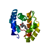
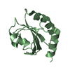
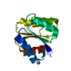
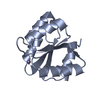
| Entry | Database: PDB / ID: 1tmy | ||||||
|---|---|---|---|---|---|---|---|
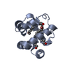
| Title | CHEY FROM THERMOTOGA MARITIMA (APO-I) | ||||||
 Components Components | CHEY PROTEIN | ||||||
 Keywords Keywords | CHEMOTAXIS / PHOSPHORYL TRANSFER / SIGNAL TRANSDUCTION | ||||||
| Function / homology |  Function and homology information Function and homology informationarchaeal or bacterial-type flagellum-dependent cell motility / phosphorelay signal transduction system / chemotaxis / metal ion binding / cytoplasm Similarity search - Function | ||||||
| Biological species |   Thermotoga maritima (bacteria) Thermotoga maritima (bacteria) | ||||||
| Method |  X-RAY DIFFRACTION / SIRAS / MOLECULAR REPLACEMENT / Resolution: 1.9 Å X-RAY DIFFRACTION / SIRAS / MOLECULAR REPLACEMENT / Resolution: 1.9 Å | ||||||
 Authors Authors | Usher, K.C. / De La Cruz, A. / Dahlquist, F.W. / Remington, S.J. | ||||||
 Citation Citation | Journal: Protein Sci. / Year: 1998 Title: Crystal structures of CheY from Thermotoga maritima do not support conventional explanations for the structural basis of enhanced thermostability. Authors: Usher, K.C. / de la Cruz, A.F. / Dahlquist, F.W. / Swanson, R.V. / Simon, M.I. / Remington, S.J. #1: Journal: J.Mol.Biol. / Year: 1994Title: Magnesium Binding to the Bacterial Chemotaxis Protein Chey Results in Large Conformational Changes Involving its Functional Surface Authors: Bellsolell, L. / Prieto, J. / Serrano, L. / Coll, M. #2: Journal: J.Mol.Biol. / Year: 1994Title: Erratum. Magnesium Binding to the Bacterial Chemotaxis Protein Chey Results in Large Conformational Changes Involving its Functional Surface Authors: Bellsolell, L. / Prieto, J. / Serrano, L. / Coll, M. #3: Journal: J.Biol.Chem. / Year: 1991Title: Crystal Structure of Escherichia Coli Chey Refined at 1.7-A Resolution Authors: Volz, K. / Matsumura, P. | ||||||
| History |
|
- Structure visualization
| Structure viewer | Molecule: MolmilJmol/JSmol |
|---|
- Downloads & links
-Download
| PDBx/mmCIF format | 1tmy.cif.gz | 34.5 KB | Display | PDBx/mmCIF format |
|---|---|---|---|---|
| PDB format | pdb1tmy.ent.gz | 23.2 KB | Display | PDB format |
| PDBx/mmJSON format | 1tmy.json.gz | Tree view | PDBx/mmJSON format | |
| Others |  Other downloads Other downloads |
-Validation report
| Arichive directory | https://data.pdbj.org/pub/pdb/validation_reports/tm/1tmyftp://data.pdbj.org/pub/pdb/validation_reports/tm/1tmy | HTTPS FTP |
|---|
-Related structure data
| Related structure data | 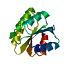 2tmyC  3tmyC 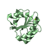 4tmyC 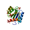 3chyS S: Starting model for refinement C: citing same article ( |
|---|---|
| Similar structure data |


-Links
 PDBj
PDBj

- Assembly
| Deposited unit | 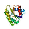
| ||||||||
|---|---|---|---|---|---|---|---|---|---|
| 1 |
| ||||||||
| Unit cell |
|
-Components
| #1: Protein | Mass: 13234.754 Da / Num. of mol.: 1 Source method: isolated from a genetically manipulated source Source: (gene. exp.) Thermotoga maritima (bacteria) / Cellular location: CYTOPLASM / Gene: CHEY / Plasmid: PQE12 / Cellular location (production host): CYTOPLASM / Gene (production host): CHEY / Production host: |
|---|---|
| #2: Water | ChemComp-HOH /  Mass: 18.015 Da / Num. of mol.: 15 / Source method: isolated from a natural source / Formula: H2O Mass: 18.015 Da / Num. of mol.: 15 / Source method: isolated from a natural source / Formula: H2O |
-Experimental details
-Experiment
| Experiment | Method: X-RAY DIFFRACTION / Number of used crystals: 1 |
|---|
- Sample preparation
| Crystal | Density Matthews: 2.2 Å3/Da / Density % sol: 50 % | ||||||||||||||||||||
|---|---|---|---|---|---|---|---|---|---|---|---|---|---|---|---|---|---|---|---|---|---|
| Crystal grow | pH: 8.5 Details: PROTEIN WAS CRYSTALLIZED FROM 0.1M TRIS BUFFER (PH 8.5) AND 1.8M AMMONIUM PHOSPHATE | ||||||||||||||||||||
| Crystal grow | *PLUS Method: vapor diffusion | ||||||||||||||||||||
| Components of the solutions | *PLUS
|
-Data collection
| Diffraction | Mean temperature: 295 K |
|---|---|
| Diffraction source | Source: ROTATING ANODE / Type: RIGAKU RUH2R / Wavelength: 1.5418 |
| Detector | Type: RIGAKU RAXIS II / Detector: IMAGE PLATE / Date: Jan 19, 1995 / Details: COLLIMATOR |
| Radiation | Monochromator: GRAPHITE(002) / Monochromatic (M) / Laue (L): M / Scattering type: x-ray |
| Radiation wavelength | Wavelength: 1.5418 Å / Relative weight: 1 |
| Reflection | Resolution: 1.9→20 Å / Num. obs: 7985 / % possible obs: 83 % / Observed criterion σ(I): 1 / Redundancy: 2.5 % / Rmerge(I) obs: 0.053 / Net I/σ(I): 5.5 |
| Reflection shell | Resolution: 1.9→2.25 Å / Redundancy: 1.5 % / Rmerge(I) obs: 0.12 / Mean I/σ(I) obs: 3 / % possible all: 69 |
- Processing
| Software |
| ||||||||||||||||||||||||||||||||||||||||||||||||||
|---|---|---|---|---|---|---|---|---|---|---|---|---|---|---|---|---|---|---|---|---|---|---|---|---|---|---|---|---|---|---|---|---|---|---|---|---|---|---|---|---|---|---|---|---|---|---|---|---|---|---|---|
| Refinement | Method to determine structure: SIRAS / MOLECULAR REPLACEMENT Starting model: PDB ENTRY 3CHY Resolution: 1.9→20 Å / Isotropic thermal model: TNT BCORREL V1.0 / σ(F): 0 / Stereochemistry target values: TNT PROTGEO Details: DISORDERED SIDE-CHAINS WERE MODELED STEREOCHEMICALLY AND HAVE THEIR OCCUPANCY SET ARBITRARILY TO 0.0.
| ||||||||||||||||||||||||||||||||||||||||||||||||||
| Solvent computation | Solvent model: BABINET SCALING / Bsol: 247 Å2 / ksol: 0.8 e/Å3 | ||||||||||||||||||||||||||||||||||||||||||||||||||
| Refinement step | Cycle: LAST / Resolution: 1.9→20 Å
| ||||||||||||||||||||||||||||||||||||||||||||||||||
| Refine LS restraints |
| ||||||||||||||||||||||||||||||||||||||||||||||||||
| Software | *PLUS Name: TNT / Version: 5E / Classification: refinement | ||||||||||||||||||||||||||||||||||||||||||||||||||
| Refinement | *PLUS Rfactor obs: 0.186 | ||||||||||||||||||||||||||||||||||||||||||||||||||
| Solvent computation | *PLUS | ||||||||||||||||||||||||||||||||||||||||||||||||||
| Displacement parameters | *PLUS | ||||||||||||||||||||||||||||||||||||||||||||||||||
| Refine LS restraints | *PLUS
|
