Resolution: 1.66→10 Å Details: SOLVENT. MOST WATER MOLECULES ARE WELL BEHAVED. A FEW WATERS WITH HIGH TEMPERATURE FACTORS AND CLOSE CONTACTS WITH OTHER WATERS MAY BE PARTIAL SITES. SOLVENT MOLECULE HOH 202 IS PROBABLY AN ...Details: SOLVENT. MOST WATER MOLECULES ARE WELL BEHAVED. A FEW WATERS WITH HIGH TEMPERATURE FACTORS AND CLOSE CONTACTS WITH OTHER WATERS MAY BE PARTIAL SITES. SOLVENT MOLECULE HOH 202 IS PROBABLY AN AMMONIUM ION. MODELLED ATOMS. THE FOLLOWING ATOMS ARE MODELS FOR THE HYPOTHETICAL BINDING POSITIONS OF MG ION AND PHOSPHORYL GROUP TO THE CHEY MOLECULE AND ARE NOT BASED UPON ELECTRON DENSITY: N ASP 57 28.900 2.872 10.690 1.00 10.00 CA ASP 57 27.660 2.298 10.194 1.00 10.00 C ASP 57 26.663 2.175 11.347 1.00 10.00 O ASP 57 27.055 1.915 12.485 1.00 10.00 CB ASP 57 27.891 0.899 9.617 1.00 10.00 CG ASP 57 26.840 0.435 8.608 1.00 10.00 OD1 ASP 57 26.936 0.853 7.123 1.00 10.00 OD2 ASP 57 25.892 -0.286 9.103 1.00 10.00 P PO3 57 27.058 -0.293 6.063 1.00 10.00 O3P PO3 57 27.145 0.067 4.652 1.00 10.00 O2P PO3 57 25.962 -1.249 6.351 1.00 10.00 O4P PO3 57 28.483 -0.924 6.407 1.00 10.00 MG MG 501 27.472 -3.123 8.987 1.00 10.00 ANOTHER POSSIBLE BINDING POSITION FOR THE PHOSPHORYL GROUP IS: N ASP 57 28.900 2.872 10.690 1.00 10.00 CA ASP 57 27.660 2.298 10.194 1.00 10.00 C ASP 57 26.663 2.175 11.347 1.00 10.00 O ASP 57 27.055 1.915 12.485 1.00 10.00 CB ASP 57 27.891 0.899 9.617 1.00 10.00 CG ASP 57 26.840 0.435 8.608 1.00 10.00 OD1 ASP 57 26.936 0.853 7.123 1.00 10.00 OD2 ASP 57 25.892 -0.286 9.103 1.00 10.00 P PO3 57 25.817 0.332 6.159 1.00 10.00 O3P PO3 57 24.535 0.863 6.679 1.00 10.00 O2P PO3 57 25.980 0.539 4.724 1.00 10.00 O4P PO3 57 25.911 -1.249 6.352 1.00 10.00
Rfactor
Num. reflection
obs
0.151
11428
Refinement step
Cycle: LAST / Resolution: 1.66→10 Å
Protein
Nucleic acid
Ligand
Solvent
Total
Num. atoms
1003
0
15
147
1165
Refine LS restraints
Refine-ID
Type
Dev ideal
Dev ideal target
X-RAY DIFFRACTION
p_bond_d
0.018
0.02
X-RAY DIFFRACTION
p_angle_d
0.049
0.04
X-RAY DIFFRACTION
p_angle_deg
X-RAY DIFFRACTION
p_planar_d
0.051
0.05
X-RAY DIFFRACTION
p_hb_or_metal_coord
X-RAY DIFFRACTION
p_mcbond_it
0.997
1
X-RAY DIFFRACTION
p_mcangle_it
1.586
1.5
X-RAY DIFFRACTION
p_scbond_it
1.455
1
X-RAY DIFFRACTION
p_scangle_it
2.452
1.5
X-RAY DIFFRACTION
p_plane_restr
0.012
0.02
X-RAY DIFFRACTION
p_chiral_restr
0.16
0.15
X-RAY DIFFRACTION
p_singtor_nbd
0.186
0.5
X-RAY DIFFRACTION
p_multtor_nbd
0.202
0.5
X-RAY DIFFRACTION
p_xhyhbond_nbd
0.224
0.5
X-RAY DIFFRACTION
p_xyhbond_nbd
X-RAY DIFFRACTION
p_planar_tor
2.3
3
X-RAY DIFFRACTION
p_staggered_tor
14.6
15
X-RAY DIFFRACTION
p_orthonormal_tor
22.1
20
X-RAY DIFFRACTION
p_transverse_tor
X-RAY DIFFRACTION
p_special_tor
+
About Yorodumi
-
News
-
Feb 9, 2022. New format data for meta-information of EMDB entries
New format data for meta-information of EMDB entries
Version 3 of the EMDB header file is now the official format.
The previous official version 1.9 will be removed from the archive.
In the structure databanks used in Yorodumi, some data are registered as the other names, "COVID-19 virus" and "2019-nCoV". Here are the details of the virus and the list of structure data.
Jan 31, 2019. EMDB accession codes are about to change! (news from PDBe EMDB page)
EMDB accession codes are about to change! (news from PDBe EMDB page)
The allocation of 4 digits for EMDB accession codes will soon come to an end. Whilst these codes will remain in use, new EMDB accession codes will include an additional digit and will expand incrementally as the available range of codes is exhausted. The current 4-digit format prefixed with “EMD-” (i.e. EMD-XXXX) will advance to a 5-digit format (i.e. EMD-XXXXX), and so on. It is currently estimated that the 4-digit codes will be depleted around Spring 2019, at which point the 5-digit format will come into force.
The EM Navigator/Yorodumi systems omit the EMD- prefix.
Related info.:Q: What is EMD? / ID/Accession-code notation in Yorodumi/EM Navigator
Yorodumi is a browser for structure data from EMDB, PDB, SASBDB, etc.
This page is also the successor to EM Navigator detail page, and also detail information page/front-end page for Omokage search.
The word "yorodu" (or yorozu) is an old Japanese word meaning "ten thousand". "mi" (miru) is to see.
Related info.:EMDB / PDB / SASBDB / Comparison of 3 databanks / Yorodumi Search / Aug 31, 2016. New EM Navigator & Yorodumi / Yorodumi Papers / Jmol/JSmol / Function and homology information / Changes in new EM Navigator and Yorodumi
 Movie
Movie Controller

 Yorodumi
 Open data
 Basic information
 Components
 Keywords
 Function and homology information
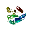
 X-RAY DIFFRACTION / Resolution: 1.66 Å
  Authors
 Citation
 Structure visualization
 Downloads & links
 Other downloads

 PDBj
PDBj

 Assembly
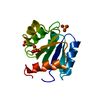

 Mass: 96.063 Da / Num. of mol.: 3 / Source method: obtained synthetically / Formula: SO4
 Mass: 18.015 Da / Num. of mol.: 147 / Source method: isolated from a natural source / Formula: H2O
 Sample preparation
 Processing