 Movie
Movie Controller

[English] 日本語
 Yorodumi
- PDB-1tid: Crystal Structures of the ADP and ATP bound forms of the Bacillus... -
+ Open data

- Basic information
| Entry | Database: PDB / ID: 1tid | ||||||
|---|---|---|---|---|---|---|---|
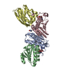
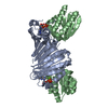
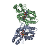
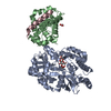
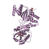
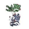
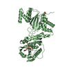
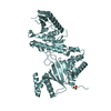
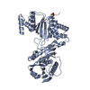
| Title | Crystal Structures of the ADP and ATP bound forms of the Bacillus Anti-sigma factor SpoIIAB in complex with the Anti-anti-sigma SpoIIAA: Poised for phosphorylation complex with ATP, crystal form I | ||||||
 Components Components |
| ||||||
 Keywords Keywords | TRANSCRIPTION / SpoIIAB / SpoIIAA / anti-sigma / anti-anti-sigma / sporulation / serine kinase | ||||||
| Function / homology |  Function and homology information Function and homology informationasexual sporulation / negative regulation of sporulation resulting in formation of a cellular spore / anti-sigma factor antagonist activity / antisigma factor binding / sigma factor antagonist activity / sporulation resulting in formation of a cellular spore / non-specific serine/threonine protein kinase / protein serine kinase activity / protein serine/threonine kinase activity / ATP binding Similarity search - Function | ||||||
| Biological species |   Geobacillus stearothermophilus (bacteria) Geobacillus stearothermophilus (bacteria) | ||||||
| Method |  X-RAY DIFFRACTION / SYNCHROTRON / MOLECULAR REPLACEMENT / Resolution: 2.5 Å X-RAY DIFFRACTION / SYNCHROTRON / MOLECULAR REPLACEMENT / Resolution: 2.5 Å | ||||||
 Authors Authors | Masuda, S. / Murakami, K.S. / Wang, S. / Olson, C.A. / Donigan, J. / Leon, F. / Darst, S.A. / Campbell, E.A. | ||||||
 Citation Citation | Journal: J.Mol.Biol. / Year: 2004 Title: Crystal Structures of the ADP and ATP Bound Forms of the Bacillus Anti-sigma Factor SpoIIAB in Complex with the Anti-anti-sigma SpoIIAA. Authors: Masuda, S. / Murakami, K.S. / Wang, S. / Olson, C.A. / Donigian, J. / Leon, F. / Darst, S.A. / Campbell, E.A. | ||||||
| History |
| ||||||
| Remark 999 | SEQUENCE THE DISCREPANCIES IN BOTH CHAINS ARE DUE TO STRAIN VARIATION. |
- Structure visualization
| Structure viewer | Molecule: MolmilJmol/JSmol |
|---|
- Downloads & links
-Download
| PDBx/mmCIF format | 1tid.cif.gz | 114.4 KB | Display | PDBx/mmCIF format |
|---|---|---|---|---|
| PDB format | pdb1tid.ent.gz | 89.2 KB | Display | PDB format |
| PDBx/mmJSON format | 1tid.json.gz | Tree view | PDBx/mmJSON format | |
| Others |  Other downloads Other downloads |
-Validation report
| Arichive directory | https://data.pdbj.org/pub/pdb/validation_reports/ti/1tidftp://data.pdbj.org/pub/pdb/validation_reports/ti/1tid | HTTPS FTP |
|---|
-Related structure data
| Related structure data |  1th8C  1thnC 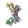 1tilC  1h4yS 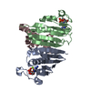 1l0oS C: citing same article ( S: Starting model for refinement |
|---|---|
| Similar structure data |

-Links
 PDBj
PDBj
- Assembly
| Deposited unit | 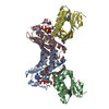
| ||||||||
|---|---|---|---|---|---|---|---|---|---|
| 1 |
| ||||||||
| Unit cell |
| ||||||||
| Details | the asymmetric unit contains one biological assembly of 2AB/2AA (a tetramer of 2 heterodimers) |
-Components
| #1: Protein | Mass: 15410.411 Da / Num. of mol.: 2 Source method: isolated from a genetically manipulated source Source: (gene. exp.) Geobacillus stearothermophilus (bacteria)Gene: SPOIIAB / Plasmid: pET21a / Species (production host): Escherichia coli / Production host: #2: Protein | Mass: 13269.778 Da / Num. of mol.: 2 / Mutation: S58A Source method: isolated from a genetically manipulated source Source: (gene. exp.) Geobacillus stearothermophilus (bacteria)Gene: SPOIIAA / Plasmid: pET21a / Species (production host): Escherichia coli / Production host: #3: Chemical |   Mass: 24.305 Da / Num. of mol.: 2 / Source method: obtained synthetically / Formula: Mg Mass: 24.305 Da / Num. of mol.: 2 / Source method: obtained synthetically / Formula: Mg#4: Chemical |   Mass: 507.181 Da / Num. of mol.: 2 / Source method: obtained synthetically / Formula: C10H16N5O13P3 / Comment: ATP, energy-carrying molecule*YM Mass: 507.181 Da / Num. of mol.: 2 / Source method: obtained synthetically / Formula: C10H16N5O13P3 / Comment: ATP, energy-carrying molecule*YM#5: Water | ChemComp-HOH / |  Mass: 18.015 Da / Num. of mol.: 62 / Source method: isolated from a natural source / Formula: H2O Mass: 18.015 Da / Num. of mol.: 62 / Source method: isolated from a natural source / Formula: H2OHas protein modification | Y | |
|---|
-Experimental details
-Experiment
| Experiment | Method: X-RAY DIFFRACTION / Number of used crystals: 1 |
|---|
- Sample preparation
| Crystal | Density Matthews: 2.87 Å3/Da / Density % sol: 57.1 % |
|---|---|
| Crystal grow | Temperature: 295.5 K / Method: vapor diffusion, hanging drop / pH: 9.7 Details: 0.1M CHESS, 2-5% DMF, 2.0 M lithium sulfate, pH 9.7, VAPOR DIFFUSION, HANGING DROP, temperature 295.5K |
-Data collection
| Diffraction | Mean temperature: 100 K |
|---|---|
| Diffraction source | Source: SYNCHROTRON / Site: NSLS  / Beamline: X25 / Wavelength: 0.9795 Å / Beamline: X25 / Wavelength: 0.9795 Å |
| Detector | Type: ADSC QUANTUM 4 / Detector: CCD / Date: Mar 8, 2002 |
| Radiation | Protocol: SINGLE WAVELENGTH / Monochromatic (M) / Laue (L): M / Scattering type: x-ray |
| Radiation wavelength | Wavelength: 0.9795 Å / Relative weight: 1 |
| Reflection | Resolution: 2.5→30 Å / Num. all: 25904 / Num. obs: 24765 / % possible obs: 95.6 % / Observed criterion σ(F): 0 / Observed criterion σ(I): 0 / Redundancy: 2.5 % / Rsym value: 0.058 / Net I/σ(I): 18.5 |
| Reflection shell | Resolution: 2.5→2.59 Å / Mean I/σ(I) obs: 5.8 / Num. unique all: 2682 / Rsym value: 0.19 / % possible all: 94.5 |
- Processing
| Software |
| |||||||||||||||||||||||||
|---|---|---|---|---|---|---|---|---|---|---|---|---|---|---|---|---|---|---|---|---|---|---|---|---|---|---|
| Refinement | Method to determine structure: MOLECULAR REPLACEMENT Starting model: pdb entry ID 1L0O, pdb entry ID 1H4Y Resolution: 2.5→30 Å / Cross valid method: THROUGHOUT / σ(F): 0 / Stereochemistry target values: Engh & Huber
| |||||||||||||||||||||||||
| Refinement step | Cycle: LAST / Resolution: 2.5→30 Å
| |||||||||||||||||||||||||
| Refine LS restraints |
|
