 Movie
Movie Controller


+ Open data

- Basic information
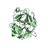
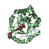
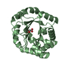
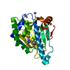
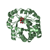
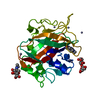
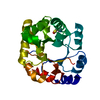
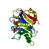
| Entry | Database: PDB / ID: 1tia | ||||||
|---|---|---|---|---|---|---|---|
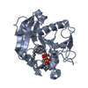
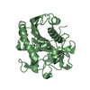
| Title | AN UNUSUAL BURIED POLAR CLUSTER IN A FAMILY OF FUNGAL LIPASES | ||||||
 Components Components | LIPASE | ||||||
 Keywords Keywords | HYDROLASE(CARBOXYLIC ESTERASE) | ||||||
| Function / homology |  Function and homology information Function and homology informationHydrolases; Acting on ester bonds; Carboxylic-ester hydrolases / lipid catabolic process / hydrolase activity Similarity search - Function | ||||||
| Biological species |  Penicillium camemberti (fungus) Penicillium camemberti (fungus) | ||||||
| Method |  X-RAY DIFFRACTION / Resolution: 2.1 Å X-RAY DIFFRACTION / Resolution: 2.1 Å | ||||||
 Authors Authors | Derewenda, U. / Swenson, L. / Yamaguchi, S. / Wei, Y. / Derewenda, Z.S. | ||||||
 Citation Citation | Journal: Nat.Struct.Biol. / Year: 1994 Title: An unusual buried polar cluster in a family of fungal lipases. Authors: Derewenda, U. / Swenson, L. / Green, R. / Wei, Y. / Dodson, G.G. / Yamaguchi, S. / Haas, M.J. / Derewenda, Z.S. #1: Journal: Protein Eng. / Year: 1994Title: Current Progress in Crystallographic Studies of New Lipases from Filamentous Fungi Authors: Derewenda, U. / Swenson, L. / Green, R. / Wei, Y. / Yamaguchi, S. / Joerger, R. / Haas, M.J. / Derewenda, Z.S. | ||||||
| History |
|
- Structure visualization
| Structure viewer | Molecule: MolmilJmol/JSmol |
|---|
- Downloads & links
-Download
| PDBx/mmCIF format | 1tia.cif.gz | 34.5 KB | Display | PDBx/mmCIF format |
|---|---|---|---|---|
| PDB format | pdb1tia.ent.gz | 11.9 KB | Display | PDB format |
| PDBx/mmJSON format | 1tia.json.gz | Tree view | PDBx/mmJSON format | |
| Others |  Other downloads Other downloads |
-Validation report
| Summary document | 1tia_validation.pdf.gz | 341.6 KB | Display | wwPDB validaton report |
|---|---|---|---|---|
| Full document | 1tia_full_validation.pdf.gz | 341.9 KB | Display | |
| Data in XML | 1tia_validation.xml.gz | 1.4 KB | Display | |
| Data in CIF | 1tia_validation.cif.gz | 3.8 KB | Display | |
| Arichive directory | https://data.pdbj.org/pub/pdb/validation_reports/ti/1tiaftp://data.pdbj.org/pub/pdb/validation_reports/ti/1tia | HTTPS FTP |
-Related structure data
| Similar structure data |
|---|
-Links
 PDBj
PDBj- Assembly
| Deposited unit | 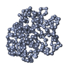
| ||||||||
|---|---|---|---|---|---|---|---|---|---|
| 1 |
| ||||||||
| Unit cell |
|
-Components
| #1: Protein | Mass: 30205.533 Da / Num. of mol.: 1 Source method: isolated from a genetically manipulated source Source: (gene. exp.) Penicillium camemberti (fungus) / References: UniProt: P61870, triacylglycerol lipase |
|---|
-Experimental details
-Experiment
| Experiment | Method: X-RAY DIFFRACTION |
|---|
- Sample preparation
| Crystal | Density Matthews: 1.93 Å3/Da / Density % sol: 36.36 % | ||||||||||||||||||||||||||||||
|---|---|---|---|---|---|---|---|---|---|---|---|---|---|---|---|---|---|---|---|---|---|---|---|---|---|---|---|---|---|---|---|
| Crystal grow | *PLUS Method: vapor diffusion, hanging drop | ||||||||||||||||||||||||||||||
| Components of the solutions | *PLUS
|
-Data collection
| Reflection | *PLUS Highest resolution: 1.98 Å / % possible obs: 79.9 % / Redundancy: 1.97 % / Rmerge(I) obs: 0.0511 |
|---|
- Processing
| Software |
| ||||||||||||||||||||||||||||||||||||||||||||||||||||||||||||||||||||||||||||||||
|---|---|---|---|---|---|---|---|---|---|---|---|---|---|---|---|---|---|---|---|---|---|---|---|---|---|---|---|---|---|---|---|---|---|---|---|---|---|---|---|---|---|---|---|---|---|---|---|---|---|---|---|---|---|---|---|---|---|---|---|---|---|---|---|---|---|---|---|---|---|---|---|---|---|---|---|---|---|---|---|---|---|
| Refinement | Resolution: 2.1→7.5 Å / σ(F): 0 /
| ||||||||||||||||||||||||||||||||||||||||||||||||||||||||||||||||||||||||||||||||
| Refinement step | Cycle: LAST / Resolution: 2.1→7.5 Å
| ||||||||||||||||||||||||||||||||||||||||||||||||||||||||||||||||||||||||||||||||
| Refine LS restraints |
| ||||||||||||||||||||||||||||||||||||||||||||||||||||||||||||||||||||||||||||||||
| Refinement | *PLUS Rfactor obs: 0.19 / Rfactor Rwork: 0.19 | ||||||||||||||||||||||||||||||||||||||||||||||||||||||||||||||||||||||||||||||||
| Solvent computation | *PLUS | ||||||||||||||||||||||||||||||||||||||||||||||||||||||||||||||||||||||||||||||||
| Displacement parameters | *PLUS | ||||||||||||||||||||||||||||||||||||||||||||||||||||||||||||||||||||||||||||||||
| Refine LS restraints | *PLUS
|
