 Movie
Movie Controller

[English] 日本語
 Yorodumi
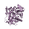
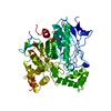
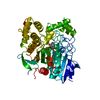
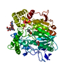
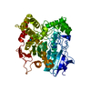
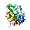
- PDB-1thg: 1.8 ANGSTROMS REFINED STRUCTURE OF THE LIPASE FROM GEOTRICHUM CANDIDUM -
+ Open data

- Basic information
| Entry | Database: PDB / ID: 1thg | ||||||||||||
|---|---|---|---|---|---|---|---|---|---|---|---|---|---|
| Title | 1.8 ANGSTROMS REFINED STRUCTURE OF THE LIPASE FROM GEOTRICHUM CANDIDUM | ||||||||||||
 Components Components | Lipase 2 | ||||||||||||
 Keywords Keywords | HYDROLASE(CARBOXYLIC ESTERASE) | ||||||||||||
| Function / homology |  Function and homology information Function and homology informationtriacylglycerol lipase / triacylglycerol lipase activity / lipid catabolic process / extracellular region Similarity search - Function | ||||||||||||
| Biological species |  Geotrichum candidum (yeast) Geotrichum candidum (yeast) | ||||||||||||
| Method |  X-RAY DIFFRACTION / Resolution: 1.8 Å X-RAY DIFFRACTION / Resolution: 1.8 Å | ||||||||||||
 Authors Authors | Schrag, J.D. / Cygler, M. | ||||||||||||
 Citation Citation | Journal: J.Mol.Biol. / Year: 1993 Title: 1.8 A refined structure of the lipase from Geotrichum candidum. Authors: Schrag, J.D. / Cygler, M. #1: Journal: J.Biol.Chem. / Year: 1993Title: Insights Into Interfacial Activation from an Open Structure of Candida Rugosa Lipase Authors: Grochulski, P. / Li, Y. / Schrag, J.D. / Bouthillier, F. / Smith, P. / Harrison, D. / Rubin, B. / Cygler, M. #2: Journal: J.Biol.Chem. / Year: 1992Title: Pancreatic Lipases: Evolutionary Intermediates in a Positional Change of Catalytic Carboxylates? Authors: Schrag, J.D. / Winkler, F.K. / Cygler, M. #3: Journal: Nature / Year: 1991Title: Ser-His-Glu Triad Forms the Catalytic Site of the Lipase from Geotrichum Candidum Authors: Schrag, J.D. / Li, Y. / Wu, S. / Cygler, M. #4: Journal: J.Mol.Biol. / Year: 1991Title: Multiple Crystal Forms of Lipases from Geotrichum Candidum Authors: Schrag, J.D. / Li, Y. / Wu, S. / Cygler, M. #5: Journal: Nature / Year: 1990Title: Structure of Human Pancreatic Lipase Authors: Winkler, F.K. / D'Arcy, A. / Hunziker, W. | ||||||||||||
| History |
|
- Structure visualization
| Structure viewer | Molecule: MolmilJmol/JSmol |
|---|
- Downloads & links
-Download
| PDBx/mmCIF format | 1thg.cif.gz | 128.9 KB | Display | PDBx/mmCIF format |
|---|---|---|---|---|
| PDB format | pdb1thg.ent.gz | 98 KB | Display | PDB format |
| PDBx/mmJSON format | 1thg.json.gz | Tree view | PDBx/mmJSON format | |
| Others |  Other downloads Other downloads |
-Validation report
| Arichive directory | https://data.pdbj.org/pub/pdb/validation_reports/th/1thgftp://data.pdbj.org/pub/pdb/validation_reports/th/1thg | HTTPS FTP |
|---|
-Related structure data
| Similar structure data |
|---|


-Links
 PDBj
PDBj


- Assembly
| Deposited unit | 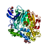
| ||||||||
|---|---|---|---|---|---|---|---|---|---|
| 1 |
| ||||||||
| Unit cell |
| ||||||||
| Atom site foot note | 1: RESIDUE 403 IS A CIS PROLINE. |
-Components
| #1: Protein | Mass: 59727.746 Da / Num. of mol.: 1 Source method: isolated from a genetically manipulated source Source: (gene. exp.) Geotrichum candidum (yeast) / Strain: ATCC 34614 / Gene: LIP2 / References: UniProt: P22394, triacylglycerol lipase |
|---|---|
| #2: Polysaccharide | 2-acetamido-2-deoxy-beta-D-glucopyranose-(1-4)-2-acetamido-2-deoxy-beta-D-glucopyranose Source method: isolated from a genetically manipulated source |
| #3: Sugar | ChemComp-NAG /   Type: D-saccharide, beta linking / Mass: 221.208 Da / Num. of mol.: 1 Type: D-saccharide, beta linking / Mass: 221.208 Da / Num. of mol.: 1Source method: isolated from a genetically manipulated source Formula: C8H15NO6 |
| #4: Sugar | ChemComp-NDG /   Type: D-saccharide, alpha linking / Mass: 221.208 Da / Num. of mol.: 1 Type: D-saccharide, alpha linking / Mass: 221.208 Da / Num. of mol.: 1Source method: isolated from a genetically manipulated source Formula: C8H15NO6 |
| #5: Water | ChemComp-HOH /  Mass: 18.015 Da / Num. of mol.: 343 / Source method: isolated from a natural source / Formula: H2O Mass: 18.015 Da / Num. of mol.: 343 / Source method: isolated from a natural source / Formula: H2O |
| Has protein modification | Y |
-Experimental details
-Experiment
| Experiment | Method: X-RAY DIFFRACTION |
|---|
- Sample preparation
| Crystal | Density Matthews: 2.3 Å3/Da / Density % sol: 46.56 % | |||||||||||||||
|---|---|---|---|---|---|---|---|---|---|---|---|---|---|---|---|---|
| Crystal grow | *PLUS pH: 6 / Method: vapor diffusion | |||||||||||||||
| Components of the solutions | *PLUS
|
-Data collection
| Radiation | Scattering type: x-ray |
|---|---|
| Radiation wavelength | Relative weight: 1 |
| Reflection | *PLUS Highest resolution: 1.8 Å / Num. obs: 48512 / % possible obs: 96.6 % / Num. measured all: 188971 / Rmerge(I) obs: 0.0393 |
- Processing
| Software |
| ||||||||||||||||||||||||||||||||||||||||||||||||||||||||||||
|---|---|---|---|---|---|---|---|---|---|---|---|---|---|---|---|---|---|---|---|---|---|---|---|---|---|---|---|---|---|---|---|---|---|---|---|---|---|---|---|---|---|---|---|---|---|---|---|---|---|---|---|---|---|---|---|---|---|---|---|---|---|
| Refinement | Rfactor Rwork: 0.157 / Rfactor obs: 0.157 / Highest resolution: 1.8 Å Details: HELICES FL1 AND FL2 FORM A FLAP COVERING THE ACTIVE SITE. THE FLAP COMPRISES RESIDUES 61 - 105. | ||||||||||||||||||||||||||||||||||||||||||||||||||||||||||||
| Refinement step | Cycle: LAST / Highest resolution: 1.8 Å
| ||||||||||||||||||||||||||||||||||||||||||||||||||||||||||||
| Refine LS restraints |
| ||||||||||||||||||||||||||||||||||||||||||||||||||||||||||||
| Refinement | *PLUS Highest resolution: 1.8 Å / Lowest resolution: 8 Å / Num. reflection obs: 43230 / σ(I): 2 / Rfactor obs: 0.157 | ||||||||||||||||||||||||||||||||||||||||||||||||||||||||||||
| Solvent computation | *PLUS | ||||||||||||||||||||||||||||||||||||||||||||||||||||||||||||
| Displacement parameters | *PLUS Biso mean: 16 Å2 | ||||||||||||||||||||||||||||||||||||||||||||||||||||||||||||
| Refine LS restraints | *PLUS
|
