 Movie
Movie Controller

[English] 日本語
 Yorodumi
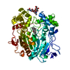
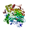
- PDB-1gz7: Crystal structure of the closed state of lipase 2 from Candida rugosa -
+ Open data

- Basic information
| Entry | Database: PDB / ID: 1gz7 | |||||||||
|---|---|---|---|---|---|---|---|---|---|---|
| Title | Crystal structure of the closed state of lipase 2 from Candida rugosa | |||||||||
 Components Components | LIPASE 2 | |||||||||
 Keywords Keywords | HYDROLASE / CARBOXYLIC ESTERASE / GLYCOPROTEIN. | |||||||||
| Function / homology |  Function and homology information Function and homology informationtriacylglycerol lipase / triacylglycerol lipase activity / lipid catabolic process Similarity search - Function | |||||||||
| Biological species |  CANDIDA RUGOSA (fungus) CANDIDA RUGOSA (fungus) | |||||||||
| Method |  X-RAY DIFFRACTION / SYNCHROTRON / MOLECULAR REPLACEMENT / Resolution: 1.97 Å X-RAY DIFFRACTION / SYNCHROTRON / MOLECULAR REPLACEMENT / Resolution: 1.97 Å | |||||||||
 Authors Authors | Mancheno, J.M. / Hermoso, J.A. | |||||||||
 Citation Citation | Journal: J.Mol.Biol. / Year: 2003 Title: Structural Insights Into the Lipase/Esterase Behavior in the Candida Rugosa Lipases Family: Crystal Structure of the Lipase 2 Isoenzyme at 1.97A Resolution Authors: Mancheno, J.M. / Pernas, M.A. / Martinez, M.J. / Ochoa, B. / Rua, M.L. / Hermoso, J.A. #1: Journal: Methods Enzymol. / Year: 1997 Title: Cloning, Sequencing, and Expression of Candida Rugosa Lipases Authors: Alberghina, L. / Lotti, M. #2: Journal: Nature / Year: 1989 Title: The Codon Cug is Read as Serine in an Asporogenic Yeast Candida Cylindracea Authors: Kawaguchi, Y. / Honda, H. / Taniguchi-Morimura, J. / Iwasaki, S. | |||||||||
| History |
|
- Structure visualization
| Structure viewer | Molecule: MolmilJmol/JSmol |
|---|
- Downloads & links
-Download
| PDBx/mmCIF format | 1gz7.cif.gz | 431.4 KB | Display | PDBx/mmCIF format |
|---|---|---|---|---|
| PDB format | pdb1gz7.ent.gz | 354.3 KB | Display | PDB format |
| PDBx/mmJSON format | 1gz7.json.gz | Tree view | PDBx/mmJSON format | |
| Others |  Other downloads Other downloads |
-Validation report
| Arichive directory | https://data.pdbj.org/pub/pdb/validation_reports/gz/1gz7ftp://data.pdbj.org/pub/pdb/validation_reports/gz/1gz7 | HTTPS FTP |
|---|
-Related structure data
| Related structure data |  1trhS S: Starting model for refinement |
|---|---|
| Similar structure data |


-Links
 PDBj
PDBj
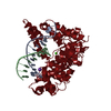
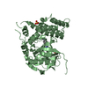
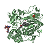
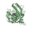
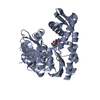
- Assembly
| Deposited unit | 
| ||||||||
|---|---|---|---|---|---|---|---|---|---|
| 1 | 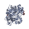
| ||||||||
| 2 | 
| ||||||||
| 3 | 
| ||||||||
| 4 | 
| ||||||||
| Unit cell |
|
-Components
| #1: Protein | Mass: 57509.344 Da / Num. of mol.: 4 / Source method: isolated from a natural source / Source: (natural) CANDIDA RUGOSA (fungus) / References: UniProt: P32946, triacylglycerol lipase#2: Polysaccharide | 2-acetamido-2-deoxy-beta-D-glucopyranose-(1-4)-2-acetamido-2-deoxy-beta-D-glucopyranose Source method: isolated from a genetically manipulated source #3: Chemical | ChemComp-GOL /   Mass: 92.094 Da / Num. of mol.: 8 / Source method: obtained synthetically / Formula: C3H8O3 Mass: 92.094 Da / Num. of mol.: 8 / Source method: obtained synthetically / Formula: C3H8O3#4: Water | ChemComp-HOH / |  Mass: 18.015 Da / Num. of mol.: 1471 / Source method: isolated from a natural source / Formula: H2O Mass: 18.015 Da / Num. of mol.: 1471 / Source method: isolated from a natural source / Formula: H2OHas protein modification | Y | |
|---|
-Experimental details
-Experiment
| Experiment | Method: X-RAY DIFFRACTION / Number of used crystals: 1 |
|---|
- Sample preparation
| Crystal | Density Matthews: 2.46 Å3/Da / Density % sol: 50 % | ||||||||||||||||||||||||
|---|---|---|---|---|---|---|---|---|---|---|---|---|---|---|---|---|---|---|---|---|---|---|---|---|---|
| Crystal grow | pH: 4.5 / Details: 15% (W/V) PEG 4000, SODIUM ACETATE 0.1M, PH 5.0 | ||||||||||||||||||||||||
| Crystal grow | *PLUS Temperature: 291 K / pH: 5 / Method: vapor diffusion, hanging drop | ||||||||||||||||||||||||
| Components of the solutions | *PLUS
|
-Data collection
| Diffraction | Mean temperature: 120 K |
|---|---|
| Diffraction source | Source: SYNCHROTRON / Site: ESRF  / Beamline: BM14 / Wavelength: 1.004 / Beamline: BM14 / Wavelength: 1.004 |
| Detector | Detector: CCD / Date: May 15, 2001 |
| Radiation | Protocol: SINGLE WAVELENGTH / Monochromatic (M) / Laue (L): M / Scattering type: x-ray |
| Radiation wavelength | Wavelength: 1.004 Å / Relative weight: 1 |
| Reflection | Resolution: 1.97→37.11 Å / Num. obs: 152503 / % possible obs: 94.6 % / Redundancy: 2 % / Biso Wilson estimate: 17.8 Å2 / Rmerge(I) obs: 0.095 / Net I/σ(I): 4.6 |
| Reflection shell | Resolution: 1.97→2.08 Å / Redundancy: 1.1 % / Rmerge(I) obs: 0.299 / Mean I/σ(I) obs: 2.2 / % possible all: 76.5 |
| Reflection | *PLUS Highest resolution: 1.97 Å / Num. measured all: 299637 / Rmerge(I) obs: 0.1 |
| Reflection shell | *PLUS % possible obs: 76.5 % / Rmerge(I) obs: 0.3 / Mean I/σ(I) obs: 2.2 |
- Processing
| Software |
| ||||||||||||||||||||||||||||||||||||||||||||||||||||||||||||
|---|---|---|---|---|---|---|---|---|---|---|---|---|---|---|---|---|---|---|---|---|---|---|---|---|---|---|---|---|---|---|---|---|---|---|---|---|---|---|---|---|---|---|---|---|---|---|---|---|---|---|---|---|---|---|---|---|---|---|---|---|---|
| Refinement | Method to determine structure: MOLECULAR REPLACEMENT Starting model: PDB ENTRY 1TRH Resolution: 1.97→11.98 Å / Rfactor Rfree error: 0.003 / Data cutoff high absF: 935285.74 / Isotropic thermal model: RESTRAINED / Cross valid method: THROUGHOUT / σ(F): 0 Details: THE ELECTRON DENSITY CORRESPONDING TO RESIDUES 78 AND 79 IS NOT CONSISTENT WITH THE SEQUENCE DESCRIBED FOR LIPASE2 (ARG-HIS), BUT WITH LEU-ASP, PRECISELY THE SEQUENCE DESCRIBED FOR LIPASE 1 AND LIPASE 3.
| ||||||||||||||||||||||||||||||||||||||||||||||||||||||||||||
| Solvent computation | Solvent model: FLAT MODEL / Bsol: 66.0866 Å2 / ksol: 0.42525 e/Å3 | ||||||||||||||||||||||||||||||||||||||||||||||||||||||||||||
| Displacement parameters | Biso mean: 27.6 Å2
| ||||||||||||||||||||||||||||||||||||||||||||||||||||||||||||
| Refine analyze |
| ||||||||||||||||||||||||||||||||||||||||||||||||||||||||||||
| Refinement step | Cycle: LAST / Resolution: 1.97→11.98 Å
| ||||||||||||||||||||||||||||||||||||||||||||||||||||||||||||
| Refine LS restraints |
| ||||||||||||||||||||||||||||||||||||||||||||||||||||||||||||
| LS refinement shell | Resolution: 1.97→2.09 Å / Rfactor Rfree error: 0.01 / Total num. of bins used: 6
| ||||||||||||||||||||||||||||||||||||||||||||||||||||||||||||
| Xplor file |
| ||||||||||||||||||||||||||||||||||||||||||||||||||||||||||||
| Refinement | *PLUS Lowest resolution: 12 Å / % reflection Rfree: 5 % / Rfactor Rfree: 0.23 / Rfactor Rwork: 0.2 | ||||||||||||||||||||||||||||||||||||||||||||||||||||||||||||
| Solvent computation | *PLUS | ||||||||||||||||||||||||||||||||||||||||||||||||||||||||||||
| Displacement parameters | *PLUS | ||||||||||||||||||||||||||||||||||||||||||||||||||||||||||||
| Refine LS restraints | *PLUS
|
